Proptosis – Feline
Rachel L. Davis1
VIN Publication
Abstract
Proptosis is sudden expulsion of the globe from the orbit causing the globe to be positioned anterior to the eyelids, with the eyelids entrapped behind the globe. Globe proptosis occurs with trauma (e.g. blunt, shearing, penetrating).
Learn about proptosis in cats, a traumatic condition where the eye is displaced from the orbit. Covers causes, diagnosis, treatment, prognosis, and emergency care options below.
Keywords: proptosis in cats, feline globe proptosis, causes of proptosis in cats, treatment for proptosis in cats, enucleation for proptosis in cats, surgical management of proptosis cats, globe displacement in cats, orbital fractures and proptosis cats, emergency treatment for eye trauma in cats, prognosis of proptosis in cats
Contributors:
1Revised by Rachel Davis DVM, MS, DACVO at Animal Eye Clinic, Westfield, Indiana, USA, on 05/04/2021
Original author was Ian P. Herring DVM, MS, DACVO, 4/18/2007
Correspondence:
Rachel L. Davis, DVM, MS, Diplomate, ACVO – Ophthalmologist
Animal Eye Clinic
4750 Killarney Drive
Carmel, IN 46033
Email: info@indyaec.com
Synonyms:
Globe proptosis
Disease Description:
Definition
Proptosis is sudden expulsion of the globe from the orbit causing the globe to be positioned anterior to the eyelids, with the eyelids entrapped behind the globe.
Etiology
Globe proptosis occurs with trauma (e.g. blunt, shearing, penetrating). A detailed history is important to determine the type of trauma sustained. The type helps to determine whether further evaluation is needed for systemic or other craniofacial trauma. In some cases, the nature of the trauma may be inferred but is not actually known. Proptosis may occur with minimal trauma in brachycephalic dogs because of their shallow orbits and large palpebral fissures. It is commonly associated with a dog fight or big dog-little dog altercation.1 Proptosis can also occur with choke injuries and overzealous restraint of brachycephalic dogs.
Orbital trauma is typically significant in mesocephalic and dolichocephalic dogs with proptosis because their orbits are larger and the globes deeper set.2 Similarly, the feline orbit is more enclosed with bone than the canine orbit, severe trauma is often required to cause proptosis.1,2
Diagnosis
Ophthalmic Examination Findings: Diagnosis is straightforward and made upon identification of the globe being located anterior to the orbit, with the eyelids entrapped behind it (Figure 1A). Concurrent mild to severe orbital or head trauma may be present, with periocular subcutaneous emphysema, soft tissue swelling, skin lacerations or palpable skull fractures. When assessing a proptosed globe, it is important to assess both the periocular structures and globe viability (see Prognosis section below). Because cats have a shallower orbit and shorter optic nerve than dogs, traction on the optic nerve may cause contralateral blindness.3 Therefore, the contralateral eye must also be assessed for vision at the time of presentation.
Physical Examination Findings: Because globe proptosis develops secondary to trauma, a thorough physical examination is performed to assess for systemic signs of trauma. In cats and non-brachycephalic dogs evaluation for concurrent facial skull fractures, subcutaneous emphysema, epistaxis, periocular tissue damage, neurologic deficits, and systemic trauma is warranted.
Other Tests: Skull and thoracic radiographs and/or computed tomography may be indicated if facial fractures or thoracic trauma are suspected. Preoperative laboratory tests are recommended if systemic trauma has occurred.
Disease Description in This Species:
Signalment
Young (mean age 4.7 yrs), unneutered male cats are at higher risk for globe proptosis, probably because of their overall increased risk of trauma secondary to territorial, roaming, and mating behavior.4 Cats are typically presented on an emergency basis, often after sustaining head trauma. Because the appearance of a proptosed globe is somewhat disturbing, most clients are distraught when they present their cat to the veterinary clinic.
Clinical Signs
The globe is positioned anterior to eyelids and orbit. The eye may be blind, with absent menace and pupillary light reflexes. Palpebral reflexes are often absent secondary to entrapment of eyelids posterior to globe. Chemosis, subconjunctival hemorrhage, bruising of the eyelids, and hyphema are all possible. The opposite eye may also have evidence of trauma (Figure 1B).
Proptosis in cats is often caused by being hit by a car and, most often, the cat leaves the area if ambulation is still possible. The cat may not return home immediately, so by the time the cat is found, the cornea and globe may be severely dried out and covered with crusty material (Figure 2). Periocular swelling, periorbital lacerations, subcutaneous emphysema, skull and mandibular fractures (Figure 3), epistaxis, and other systemic signs of trauma may be present. Severe head trauma and alterations in mentation and other cranial nerves may be noted.
Etiology:
Trauma
Breed / Species Predilection:
None, no breed signalment
Sex Predilection:
Male intact
Age Predilection:
Young adult
| Diagnostic Procedures: | Diagnostic Results: | |
| Radiography of head/skull | Facial fracture | |
| Computed tomography | Skull fracture/s | |
| (superior to radiography) Ocular examination | Cataract, lens opacity | |
| Corneal fluorescein staining positive | ||
| Eyelid, conjunctival laceration | ||
| Hyphema, blood anterior chamber eye | ||
| RETINAL CHANGES | ||
| Retinal detachment | ||
| Retinal hemorrhages | ||
| Retinal pigment abnormal | ||
| Vitreous cloudy |
Images:
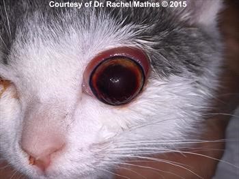
Figure 1A. A young cat with a left globe proptosis. Notice the dark hyphema in the anterior chamber and ipsilateral epistaxis as a result of the trauma (hit-by-car). An irregular flash artifact on the corneal surface is secondary to corneal drying and exposure. 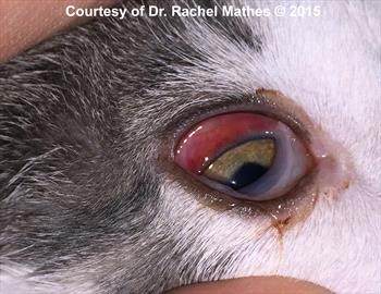
Figure 1B. The opposite eye (OD) of the same cat. Subconjunctival hemorrhage is present that will resolve spontaneously over 3-5 days. This eye is visual. 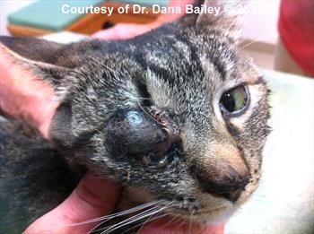
Figure 2. Dried-out proptosed eye.
Click here to see board discussion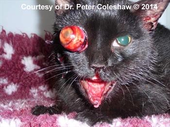
Figure 3. Proptosis OD and mandibular degloving injury (photograph)
Click here to see board discussion
Treatment / Management:
SPECIFIC THERAPY
Prognosis for vision in cats with proptosed globes is grave and other significant craniofacial injuries are often sustained in these cases.2,4 Surgery on the eye must often be delayed for several days in cats with severe head and systemic injuries.
Enucleation
Enucleation is usually required for proptosis in the cat. Enucleation is recommended if significant orbital or facial trauma is present; if the globe is severely dried or damaged; or if surgery must be delayed. Although proptosis has not been linked to feline post-traumatic ocular sarcoma, enucleation should also be considered in young cats with concurrent intraocular lens damage to avoid the potential risk of tumor development later in life.5,6 Care must be taken during enucleation to prevent further traction on the optic nerve and subsequent contralateral blindness. Ligation of the optic nerve in cats must be done very carefully (e.g. avoid traction on the nerve, gentle placement of hemoclips) or avoided altogether for this reason.3
Surgical Reduction and Tarsorrhaphy
Reduction of a proptosed feline globe may be viewed as a cosmetic procedure since studies have not shown any cat with a proptosis to have been reported to regain vision. Reference 2. Surgery to reduce the globe should only be pursued if there is a high degree of certainty that the periocular and orbital structures are healthy. Because cats with craniofacial trauma often have maxillary and orbital fractures, orbital integrity must be assessed (ideally with computed tomography) prior to reduction of a proptosed globe. For more information on reduction of a proptosed globe, see the Canine Associate chapter on Proptosis.
SUPPORTIVE THERAPY
Supportive therapy of craniofacial and/or systemic trauma is instituted as needed. Intravenous fluid therapy, analgesics, and antibiotics are administered as indicated. Topical ocular lubricants and/or antibiotics are used to protect the globe until surgery can be performed.
MONITORING and PROGNOSIS
After enucleation surgery re-evaluate the cat in 10-14 days to ensure that normal healing has occurred. Follow up for other craniofacial and systemic trauma is based on the specific therapy required and the nature of the trauma. Recommend castration of any intact male cat and/or keeping the cat inside in order to minimize future traumatic injuries.
After globe reduction, the patient is re-evaluated in 5-7 days. If the globe has returned to a normal position and all eyelid/periocular swelling has resolved or if complications are occurring (e.g. purulent discharge, persistent pain), the central tarsorrhaphy suture may be removed. Ideally, the tarsorrhaphy should remain in place for 10-14 days to allow complete healing of the periocular and orbital tissues. Prognosis for return of vision is grave.
Special Considerations:
Other Resources:
VIN Message Board discussions on proptosis
Client Handout on eye injuries
Proceedings articles that discuss proptosis
Differential Diagnosis:
Exophthalmos
Orbital fractures pathology, IOP monitoring should be performed periodically (q2-6mo depending on the severity of disease). This is especially because glaucoma in cats is not as clinically apparent to the owner or clinician compared to dogs.
References:
- Samuelson D A, Gelatt K N : Ophthalmic Anatomy. Veterinary Ophthalmology, 4th ed. Wiley-Blackwell, Oxford UK pp. 38-40.
- Gilger BC, Hamilton HL, Wilkie DA, et al: Traumatic ocular proptoses in dogs and cats: 84 cases (1980-1993). J Am Vet Med Assoc 1995 Vol 206 pp. 1186-1190.
- Donaldson D, Riera M M, Hlloway A, et al: Contralateral optic neuropathy and retinopathy associated with visual and afferent pupillomotor dysfunction following enucleation in six cats. Vet Ophthalmol 2014 Vol 17 (5) pp. 373-84.
- Fritsche J, Ruhli M, Spiess B, et al: [Prolapse of the eyeball in small animals: a retrospective study of 36 cases]. Tierarztl Prax (German) 1996 Vol 24 pp. 55-61.
- Dubielzig RR, Everitt J, Shadduck JA, et al: Clinical and morphologic features of post-traumatic ocular sarcomas in cats. Vet Pathol 1990 Vol 27 pp. 62-65.
- Zeiss CJ, Johnson EM, Dubielzig RR: Feline intraocular tumors may arise from transformation of lens epithelium. Vet Pathol 2003 Vol 40 pp. 355-362.
- Mandell DC: Ophthalmic emergencies. Clin Tech Small Anim Pract 2000 Vol 15 (2) pp. 94-100.
- Cho J: Surgery of the globe and orbit. Top Companion Anim Med 2008 Vol 23 (1) pp. 23-37.
- Giuliano E A: Feline ocular emergencies. Clin Tech Small Anim Pract 2005 Vol 20 (2) pp. 135-41.