A novel modified intradermal marginal closure for blepharoplasty in dogs: description and evaluation of post-surgical outcome in 146 eyes
Rachel L. Davis1 | CA Creek2 | PA Moore2 | KE Myrna2
Abstract
Purpose
To describe and evaluate a novel eyelid marginal closure.
Methods
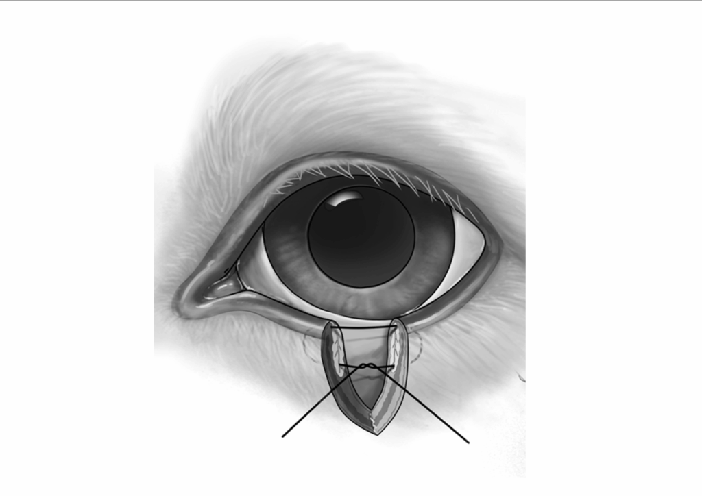
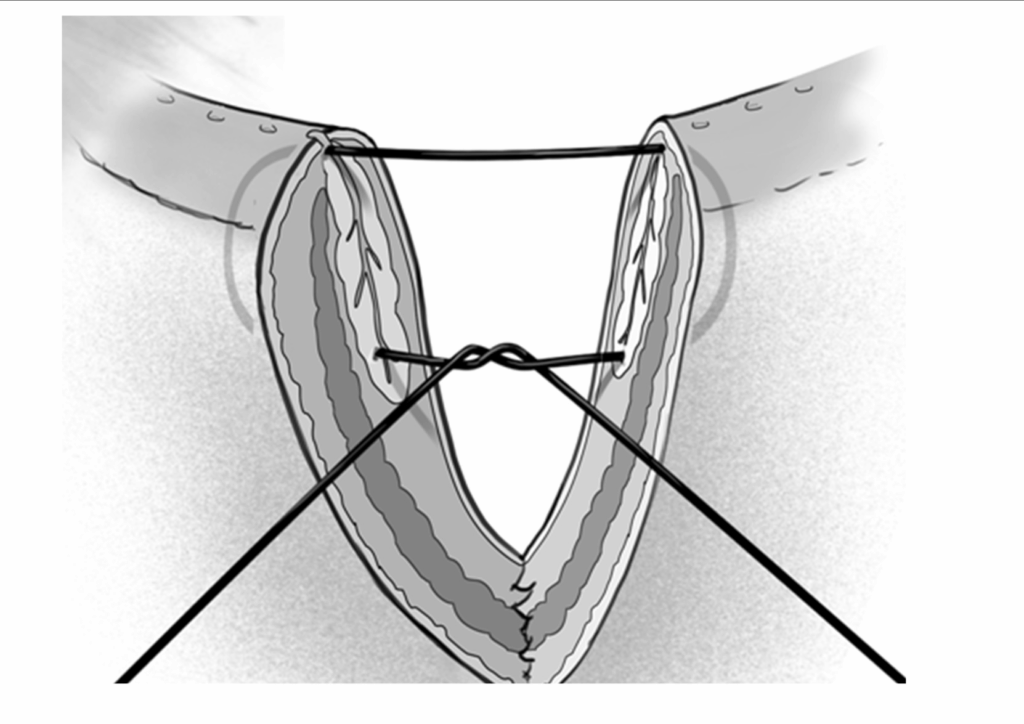
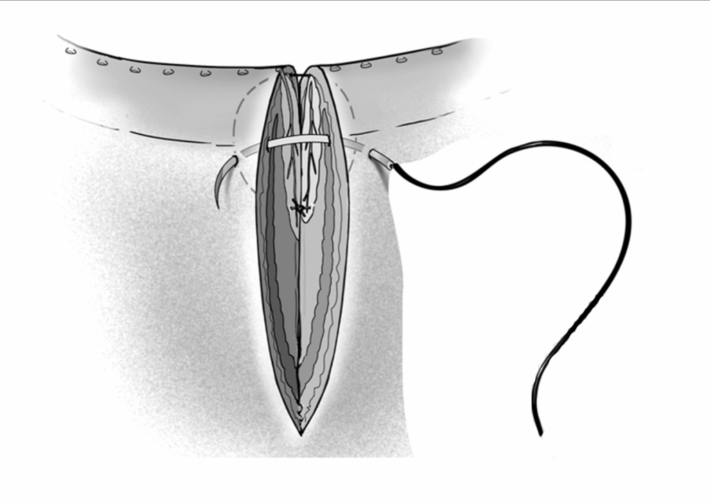
Records from dogs undergoing blepharoplasty at the University of Georgia VTH from January 2008 to February 2012 were reviewed. All patients included had a full-thickness marginal defect closed with a single simple interrupted absorbable 6–0 intradermal suture originating at the base of the cut Meibomian gland to exit the dermis at the level of the eyelid margin, passed back through the contiguous cut lid interface in a mirrored fashion and tied to create marginal apposition with a buried knot. A two-layer closure was completed with simple interrupted skin sutures. Breed, sex, age, marginal apposition, intra- and post-operative complications, follow up time and post-operative outcome were recorded.
Results
The described closure was performed on eighty two dogs (146 eyes) representing twenty four breeds for marginal eyelid mass removal (14), entropion correction (84), medial canthoplasty (18) or lateral canthal closure (30). Follow up (0.5 – 31 months) was available for 143 eyes. All closures were successful with no suture-related corneal ulcers, no lid distortion, no dehiscence and no appositional failure. Advantages of this procedure include no eyelid margin suture contact, superior marginal stability, ease of execution and application to thin eyelids.
Conclusions
A novel simple marginal eyelid closure is described with excellent postoperative. Results: This technique is easy to execute, attains ideal marginal apposition, has application to numerous adnexal surgeries and causes no corneal irritation, indicating that it may be superior to figure-of-eight and U-form pattern marginal closures. None.
Images

Keywords: canine, blepharoplasty dogs, intradermal marginal closure dogs, eyelid surgery dogs, canine eyelid closure technique, novel blepharoplasty method dogs, eyelid margin reconstruction dogs, post-surgical outcomes eyelid surgery dogs, veterinary ophthalmology blepharoplasty, entropion correction dogs, eyelid mass removal dogs
1Animal Eye Clinic, Westfield, Indiana, USA
2Department of Small Animal Medicine and Surgery, College of Veterinary Medicine, University of Georgia, Athens, GA, USA
Correspondence:
Rachel L. Davis, DVM, MS, Diplomate, ACVO – Ophthalmologist
Animal Eye Clinic
4750 Killarney Drive
Carmel, IN 46033
Email: info@indyaec.com