Concurrent clinical intraocular findings in horses with depigmented punctate chorioretinal foci
Rachel L. Davis1,2 | Erin L. Burdette2 | Phillip A. Moore2 | Kathern E. Myrna2
Abstract
Objective
To report concurrent clinical intraocular findings in horses with depigmented punctate chorioretinal foci and to document any correlation with equine recurrent uveitis (ERU).
Procedure
Records of 131 horses (241 eyes) examined at the University of Georgia Veterinary Teaching hospital from 2001 to 2010 were reviewed with either clinically normal fundi or depigmented punctate chorioretinal foci in the absence of other fundic pathology. Data collected included patient signalment, concurrent clinical ocular findings and follow-up information. Sex, presence of no other intraocular findings, presence of ERU, presence of cataracts, and presence of vitreal disease were compared between normal and foci groups using chi-squared analysis. Age and length of follow-up time were compared using a student’s t-test.
Results
Ninety-one horses (167 eyes) with chorioretinal foci and forty horses (74 eyes) with clinically normal ocular fundi were examined. Fifty-eight (64%) horses with chorioretinal foci and 20 (50%) horses with clinically normal fundi had a normal intraocular examination. There was no significant difference in any of the criteria examined between groups.
Conclusions
Horses with depigmented punctate chorioretinal foci, in the absence of other fundic pathology, are not more likely to have intraocular disease or ERU than horses with clinically normal ocular fundi. These findings suggest that depigmented punctate fundic foci in horses are not indicative of or associated with ERU.
Images
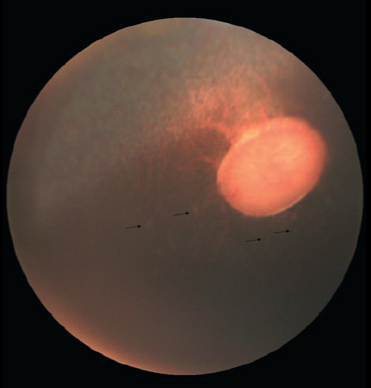
A clinical fundic photograph of the non-tapetal region and optic nerve head of a representative horse (8 year-old gelding Warmblood) in this study is depicted. The chorioretinal foci are punctate, whitish-gray, and flat (arrows). In our study, they were most often observed in the peripapillary non-tapetal region, as depicted here, although they were also observed in the tapetal and peripheral non-tapetal regions. Two flash artifacts are present over the optic nerve head in this photograph. 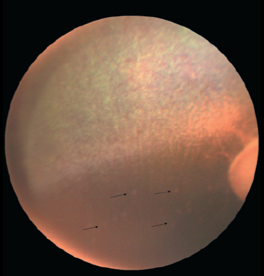
A clinical fundic photograph (8 year-old gelding Warmblood) of the peripheral tapetal and non-tapetal region is depicted with the optic nerve head pictured at the far right. The chorioretinal foci are punctate, whitish-gray, and flat (arrows). The foci depicted here affect the peripapillary region and extend into the peripheral non-tapetal region. 
Presence of no other intraocular findings, presence of
equine recurrent uveitis, presence of cataracts, and presence of vitreal
disease were compared between horses with chorioretinal foci and
horses without chorioretinal foci. There was no significant difference
between groups in any criteria evaluated.
Keywords: equine ophthalmology, depigmented chorioretinal foci, punctate fundic lesions horses, equine recurrent uveitis ERU, equine fundus exam, horse retina depigmentation, intraocular findings horses, chorioretinal scars horses, equine retinal disease, veterinary ophthalmology horses, equine eye, horse retina, horse eye disease, equine fundus, ERU horse, horse vision, eye lesions horse, retina spots horse, horse uveitis, equine ophthalmology
1Animal Eye Clinic, Westfield, Indiana, USA
2Department of Small Animal Medicine and Surgery, College of Veterinary Medicine, University of Georgia, Athens, GA 30602, USA
Correspondence:
Rachel L. Davis, DVM, MS, Diplomate, ACVO – Ophthalmologist
Animal Eye Clinic
4750 Killarney Drive
Carmel, IN 46033
Email: info@indyaec.com