Published by Emily A. Latham, DVM
Contributors:
Revised by Emily A. Latham MVB and Rachel L. Davis DVM, MS, DACVO
Revised by Andrew Lewin BVM&S, MRCVS, DACVO, 4/2/2019
Original author was Ralph E. Hamor DVM, MS, DACVO, 9/27/2012
Synonyms:
Cataract-associated uveitis
Lens-induced iridocyclitis
Lens-induced iritis
Phacolytic uveitis
Disease Description:
Definitions
Lens-induced uveitis (LIU) is intraocular inflammation affecting the anterior uveal tract that is associated with the release of lens proteins into the anterior chamber. In animals, LIU is divided into two types. Phacolytic uveitis is associated with cataract formation, especially rapidly developing or progressing or longstanding, hypermature cataracts.1,2 It occurs secondary to leakage of soluble lens proteins through an intact lens capsule. When the term, LIU, is used most clinicians are referring to this form of uveitis.
Phacoclastic uveitis is generally a more severe anterior uveitis that is associated with traumatic or spontaneous lens capsule rupture. It can result in vision-threatening intraocular inflammation that requires aggressive medical and/or surgical therapy.1,3-5
Etiology and Pathophysiology
Lens-induced uveitis is caused by the release of lens proteins through the lens capsule from a cataractous lens.1,6 Embryologically, the lens placode forms from ectoderm, whereas other ocular structures form from mesoderm. The lens and its proteins are sequestered from mesodermal tissue by the lens capsule. Leakage of lens proteins triggers an immune-mediated response because the proteins are recognized as “foreign”. The immune-mediated reaction causes secondary intraocular inflammation. Phacolytic uveitis is uncommon in cats because primary, inherited cataracts are rare.12
Most cataracts in cats develop secondary to uveitis, and uveitis typically develops first.12 Cataracts caused by uveitis generally begin in the cortex and are slow to progress.13 The stage of cataract development; presence of other ocular or systemic abnormalities; and severity of the uveitis help to determine whether the inflammation is secondary to the cataract. Typically, LIU is milder than uveitis from other causes.11 LIU is unlikely with incipient cataracts; variable with immature and mature cataracts; and most likely with hypermature cataracts. By definition, a hypermature cataract is one that is changing size and/or shape, usually from leakage of lens proteins.
Diagnosis
Physical Examination Findings/History: A cataract is present in the affected eye(s), along with clinical evidence of ocular inflammation. There may be no history of prior uveitis or systemic disease that can cause uveitis. The rest of the physical examination may be normal, or any abnormalities may reflect the inciting cause of the cataract.
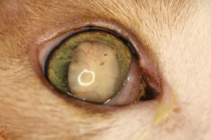
Ocular Examination Findings: Evidence of anterior uveitis (Figure) is present, such as episcleral congestion, corneal edema, perilimbal corneal neovascularization, keratic precipitates, aqueous flare, hypopyon, hyphema, fibrin in the aqueous humor, miosis, dyscoria, debris, vascularization, and/or pigment on the anterior lens capsule, wrinkling of the lens capsule, and discoloration, thickening or hyperemia (rubeosis iridis) of the iris. If the posterior segment can be visualized, vitreal haze, vitreal liquefaction/degeneration, retinal detachment, and retinal hemorrhages may be present. Measurement of IOP is important in all cases of LIU. IOP can be low (acute anterior uveitis), normal, or elevated (secondary glaucoma).
Laboratory Tests: If doubt exists as to the cause of the cataract or uveitis, and no other intraocular causes are apparent, a complete work-up for a systemic disease should be considered. See the Feline VINcyclopedia chapters on Uveitis and Cataracts for recommendations.
Ultrasonography: Ultrasonography can be used to rule out posterior segment disease when the vitreous and retina cannot be visualized through the cataract. Ultrasonography can also be used to identify a lens capsule rupture to rule out phacoclastic uveitis.12
Disease Description in This Species:
Signalment
No breed, sex, or age predispositions have been noted.
Clinical Signs
In mild cases the only clinical signs may be conjunctival/episcleral hyperemia and cataract formation that are not recognized by the owner. LIU is more likely to be chronic than other forms of uveitis. Common signs with moderate to severe LIU include visual deficits, epiphora, blepharospasm, photophobia, conjunctival hyperemia, episcleral congestion, corneal edema, perilimbal corneal neovascularization, and other evidence of anterior uveitis.
Etiology:
Cataract
Breed / Species Predilection:
None
Sex Predilection:
None
Age Predilection:
None
Clinical Findings:
- AFEBRILE
- Anisocoria, pupils unequal
- Anterior lens capsule debris and/or pigment deposition
- Blepharospasm, eye pain
- BLINDNESS OR OTHER VISUAL DEFICIT
- Blindness partial, visual deficit
- Conjunctival congestion, hyperemia
- CONJUNCTIVITIS
- Corneal vascularization
- Edema conjunctiva, chemosis
- Edema corneal
- EDEMA or SWELLING
- Enophthalmos
- Epiphora, lacrimation increased
- Episcleral injection/congestion
- Glaucoma
- HEMORRHAGE
- Hypopyon
- Iris bombe
- Iris depigmentation
- Iris pigmentation abnormal
- Keratic precipitates
- Keratoconjunctivitis
- Lens luxation, subluxation
- Miosis
- OCULAR DISCHARGE
- PAIN
- PHOTOPHOBIA
- Phthisis bulbi
- Rubeosis iridis, iris hyperemia
- Synechia, anterior
- Third eyelid, nictitating membrane prolapsed
- UVEITIS
- Uveitis, anterior
- ZZZ INDEX ZZZ
| Diagnostic Procedures: | Diagnostic Results: | |
| Ocular examination | Anterior chamber deep eye | |
| Aqueous flare | ||
| Cataract, lens opacity | ||
| Hyphema, blood anterior chamber eye | ||
| Intraocular pressure increased on tonometry | ||
| Intraocular pressure normal or decreased on tonometry if uveitis present | ||
| Lens absent, small or deformed | ||
| Leukocoria, leukokoria | ||
| Preiridal fibrovascular membrane | ||
| Vitreous cloudy |
Images:
Figure. Chronic lens-induced uveitis

17-yr-old cats. Cataract is hypermature. Evidence of longstanding (posterior synechiae, dyscoria, lens capsule vascularization and pigmentation), low-grade (no aqueous flare or keratitic precipitates) uveitis is present.
Treatment / Management:
The goal of therapy is to control pain and intraocular inflammation to decrease the chance of secondary glaucoma and other potential sequelae. If inflammation can be controlled so lensectomy can be pursued, then restoration of vision is also an objective.
SPECIFIC THERAPY
Medical Therapy
Topical Corticosteroids: Topical steroids are indicated if LIU is severe, with aqueous flare; swelling/discoloration of the iris; aqueous humor fibrin; developing posterior synechiae, etc. Prednisolone acetate 1% or dexamethasone 0.1% may be administered initially q 4-6 hrs, then reduced as LIU is controlled. Topical corticosteroids have potential side effects, including increased risk of infection; potentiation of collagenase activity that may lead to melting of an ulcerated cornea; and delayed wound and ulcer healing. Topical steroids are also absorbed systemically. Steroid use can reactivate latent feline herpesvirus (FHV-1) infections.12 It may be prudent to also begin anti-viral medications in cats with a history of prior clinical FHV-1 infections.12
Topical Nonsteroidal Anti-Inflammatory Drugs (NSAIDS): NSAIDs can be applied q 6-24 hrs. Examples include flurbiprofen 0.03% and diclofenac 0.1%. NSAIDS are typically used in situations where topical steroids are contraindicated; LIU is low grade; and/or chronic therapy is required. Topical NSAIDs are avoided or used judiciously if glaucoma is present (or impending) because they can increase IOP. Because topical NSAIDs may also delay corneal healing, avoid their use when corneal ulceration is present.7
Systemic Anti-inflammatory Drugs: Systemic corticosteroids may also be administered for severe LIU when no underlying disease is present that contraindicates their use. Systemic NSAIDs may be preferred in certain medical situations, and the two classes of drugs are never administered concurrently. Potential side effects of long-term systemic steroids and NSAIDs are well documented and must be taken into consideration.
Surgical Therapy
Surgical therapy is removal of the cataract (lensectomy). However, eyes with severe LIU may not be candidates for surgery. In eyes that have experienced LIU, postoperative complication rates are higher (e.g. increased uveitis, glaucoma, retinal detachment), and success rates are often lower.11 Very few reports exist of long-term success rates for surgical lens extraction in cats with LIU.3,7
SUPPORTIVE THERAPY
Mydriatics/cycloplegics may be applied q 12-48 hrs to prevent or decrease the risk of posterior synechiae formation; provide analgesia by eliminating ciliary body spasm (cycloplegia); and stabilize the blood-aqueous barrier. Atropine is generally given to effect and is tapered as the uveitis is controlled to the lowest frequency that maintains mydriasis and pain control. Monitoring IOP is essential when using mydriatics to treat uveitis because secondary glaucoma may develop. Mydriatics are not used if glaucoma is present or suspected. With ciliary body inflammation, IOP is typically low with active uveitis. Therefore, an eye with significant uveitis and normal IOP may be in the early stages of secondary glaucoma. The use of atropine must be carefully monitored or limited in these cases. The ointment form of atropine is preferred in cat because liquid formulations may cause profuse hypersalivation and distress.
If glaucoma is present, medical therapy with topical beta blockers (e.g. timolol) and/or carbonic anhydrase inhibitors (e.g. dorzolamide) is started. Enucleation may be warranted for eyes that are blind, unresponsive to therapy, chronically inflamed, and/or painful.
MONITORING
Frequent, chronic monitoring is essential to evaluate response to therapy; monitor progression of the disease; and detect complications. Because IOP can change over time, it is commonly measured at each recheck examination. It is important to note that hypotonic (low IOP) eyes may not become normotensive again, even after all inflammation is clinically controlled. Hence, low IOP measurements cannot be used alone to determine whether continued anti-inflammatory therapy is needed. Long-term or permanent therapy may be required for LIU.
PROGNOSIS
Prognosis varies, depending on the stage and progression of the cataract; presence of complications at the time of diagnosis; ability to control uveitis; and whether an opportunity arises to remove the cataract. LIU in some eyes can be controlled with appropriate anti-inflammatory drugs and careful monitoring. Medical therapy does not cure LIU as long as leakage of lens proteins continue from the cataract.12
Prolonged LIU can lead to corneal edema, dyscoria, peripheral anterior synechiae, ectropion uvea, entropion uvea, posterior synechiae, iris bombé, hypo- or hyperpigmentation of the iris, iris atrophy, pre-iridal fibrovascular membrane formation, secondary glaucoma, lens subluxation/luxation, vitreal degeneration (liquefaction/syneresis), vitreal traction bands, retinal degeneration, retinal detachment, chorioretinal scarring, endophthalmitis, panophthalmitis, phthisis bulbi, and permanent blindness.
Preventive Measures:
Animals with inherited cataracts should not be bred. Surgical removal of cataracts can often prevent development of LIU when performed before the cataract becomes mature or hypermature. Some clinicians advocate prophylactic use of topical NSAIDs in cataractous eyes to try and prevent development of LIU but clinical studies on the efficacy of prophylactic therapy are lacking.
Differential Diagnosis:
Other causes of uveitis
Phacoclastic uveitis
References:
- Fischer C: Lens-induced uveitis. Comparative Ophthalmic Pathology Wiley-Blackwell 1983 pp. 254-63.
- Wilcock BP, Peiffer RL: The Pathology of Lens-induced Uveitis in Dogs. Vet Pathol 1978 Vol 24 (6) pp. 549-43.
- Braus BK, Tichy A, Featherstone HJ, et al: Outcome of phacoemulsification following corneal and lens laceration in cats and dogs (2000-2010). Vet Ophthalmol 2017 Vol 20 (1) pp. 4-10.
- Paulsen ME, Kass PH: Traumatic corneal laceration with associated lens capsule disruption: a retrospective study of 77 clinical cases from 1999 to 2009. Vet Ophthalmol 2012 Vol 15 (6) pp. 355-68.
- Pont RT, Riera MM, Newton R, et al: Corneal and anterior segment foreign body trauma in dogs: a review of 218 cases. Vet Ophthalmol 2016 Vol 19 (5) pp. 386-97.
- Ofri R: Lens. In: Maggs DJ, Miller PE, Ofri R (eds): Slatter’s Fundamentals of Veterinary Ophthalmology, 4th ed. Elsevier Saunders, St. Louis 2008 pp. 263-64.
- Benz P, Maass G, Csokai J, et al: Detection of Encephalitozoon cuniculi in the feline cataractous lens. Vet Opthalmol 2011 Vol 14 (1) pp. 37-47.
- Hersh PS, Rice BA, Baer JC, et al: Topical nonsteroidal agents and corneal wound healing. Arch Ophthalmol 1990 Vol 108 (4) pp. 577-83.
- Van Der Woerdt A: Lens-induced uveitis. Vet Ophthalmol 2000 Vol 3 (4) pp. 227-234.
- Dietz HH, Jensen OA, Wissler J: Lens-Induced Uveitis in a Domestic Cat. Nordisk Veterinaermedicin 1985 Vol 37 (1) pp. 10-15.
- Van Der Woerdt A, Nasisse MP, Davidson MG: Lens-induced uveitis in dogs: 151 cases (1985-1990). J Am Vet Med Assoc 1992 Vol 201 (6) pp. 921-6.
- xxx: xxx. In: Gelatt KN, Hendrix DVH, Ben-Shlomo G, et al (eds): Veterinary Ophthalmology, 6th ed. Wiley-Blackwell, Ames IA 2021.
- Guyonnet A, Donzel E, Bourguet A, et al: Epidemiology and clinical presentation of feline cataracts in France: A retrospective study of 268 cases. Vet Ophthalmol 2019 Vol 22 (2) pp. 116-24.
Feedback:
If you note any error or omission or if you know of any new information, please send your feedback to VINcyclopedia@vin.com.
If you have any questions about a specific case or about this disease, please post your inquiry to the appropriate message boards on VIN.
