Publication: Veterinary Information Network (VIN)
Disease Description
Uveal melanoma is the most common primary intraocular tumor in cats typically seen as an intraocular pigmented mass (or masses).1,2 While most uveal melanomas are pigmented, amelanotic tumors are possible. Unlike the usually benign tumor in dogs, this tumor tends to be metastatic in cats, with a metastatic rate of 60-70% reported in the literature.3 Two clinical variations of the disease may be seen with one type presenting as a focal, pigmented mass arising from the iris, ciliary body or choroid and the other type presenting as flat, pigmented, typically multifocal areas of progressive iris pigmentation (see iris melanosis). There may be some cross-over between the clinical variations of feline uveal melanoma with the flat lesions becoming raised and nodular later in the disease process (figure). Additionally, as the mass grows, fills the eye and variably distorts or destroys the intraocular structures, differentiation between an originally flat, irial mass or raised, solitary mass is clinically unapparent and irrelevant. For the purposes of this discussion, focal, solitary uveal masses or pigmented masses filling and distorting intraocular structures will be discussed here while flat, iridal pigmented lesions, also termed iris melanosis, will be discussed elsewhere.
Etiology
Uveal melanomas arise as primary, intraocular tumors in cats typically arising from the iris or ciliary body.3,4 Primary choroidal intraocular melanomas have been reported, but are considered rare.5 Almost all feline melanomas are considered malignant based on histopathologic grading, but not all may metastasize. Because the metastatic rate is high; however, identification of these tumors with appropriate treatment (usually enucleation) is critical.6 A relationship between feline sarcoma virus and uveal melanoma was suggested in early reports,7 but later studies showed any lack of relationship between uveal melanoma formation and either feline sarcoma viruses or feline leukemia virus.8
Diagnosis
Ophthalmic Examination Findings
The initial clinical presentation of uveal melanomas in cats is widely varied, depending on the location and growth rate. Early tumors typically appear as raised, focal, brown or black intrairidal masses within the iris body or at the iris base, especially if emerging anteriorly from a primary ciliary body melanoma. Depending on the growth rate, these masses may be relatively well-demarcated9 or, more commonly, poorly demarcated, irregular tumors.4 Brown or black intrairidal masses should be differentiated from iris cysts based on transillumination or ocular ultrasound. Choroidal melanomas may appear as black masses visible on fundic examination and emerging into the vitreous.5
Later, tumors may appear as large, pigmented intraocular tumors with variable intraocular involvement. Large, invasive uveal melanomas may cause dyscoria, secondary glaucoma, anterior uveitis, hyphema, buphthalmos, lens subluxation, ocular pain, retinal detachments or blindness.3,10 These large, invasive tumors may be very aggressive in cats with rapid local expansion and distant metastasis.10 Depending on the stage of glaucoma and whether uveitis or hyphema is present, it may not be clinically apparent on presentation that an intraocular tumor is present based on ophthalmic examination alone.
Physical Examination Findings
Because feline uveal melanomas are typically malignant with a high metastatic rate, full systemic staging is recommended for cats presenting with pigmented, intraocular tumors. These tumors tend to metastasize to the lymph nodes, lungs and liver,3 although any visceral organ may be affected and skeletal metastasis has been reported.11 Thorough physical examination is warranted with a client discussion regarding further diagnostics.
Disease Description in this Species
Signalment
Uveal melanoma is typically seen in middle-aged to older cats, with a reported mean age of 96-11yr.3 No breed predisposition has been reported although most affected cats in studies have been domestic.
Clinical Signs
Early tumors typically appear as raised, focal, brown or black intrairidal masses within the iris body or at the iris base, especially if emerging anteriorly from a primary ciliary body melanoma. Depending on the growth rate, these masses may be relatively well-demarcated7 or, more commonly, poorly demarcated, irregular tumors.4 A solitary, well-demarcated pigmented iridal mass in a cat that was monitored and did not metastasize for at least a year was documented; however, this presentation is rare in cats.9 If monitoring of focal iris masses in cats is pursued, this should be done with caution and frequent follow up. Later, tumors may appear as large, pigmented intraocular tumors with variable intraocular involvement. Large, invasive uveal melanomas may cause dyscoria, secondary glaucoma, anterior uveitis, hyphema, buphthalmos, lens subluxation, ocular pain, retinal detachments or blindness.3,10 These large, invasive tumors may be very aggressive in cats with rapid local expansion and distant metastasis.10 Depending on the stage of glaucoma and whether uveitis or hyphema is present, it may not be clinically apparent on presentation that an intraocular tumor is present based on ophthalmic examination alone.
Etiology
- Spontaneous
- Neoplastic
Breed Predilection
- None
Sex Predilection
- None
Age Predilection
- Mean age 11yrs
Diagnostic Procedures
Any larger, solid, pigmented intraocular mass distorting the iris, anteriorly displacing the iris or displacing other intraocular structures may be presumed to be a uveal melanoma, although histopathology is required for definitively diagnosis. Uveal melanomas may emerge through the sclera to affect the perilimbal region, thus making it difficult to distinguish between a primary limbal melanoma and a primary uveal melanoma.1 Ocular ultrasound and gonioscopy will help determine the extent of the mass and involvement with intraocular structures. These diagnostics will also help distinguish a primary limbal melanoma vs. uveal melanoma.
Images

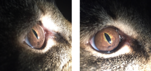
A 12yr old FS DSH is depicted with extensive iridal pigmentation (see iris melanosis) and ventral, lobulated, pigmented, iridal masses. Systemic staging showed no evidence of metastasis at the time of presentation and an enucleation was performed. Malignant iris melanoma was diagnosed and serial thoracic radiographs were recommended at six and twelve months after surgery.
Treatment/Management/Prognosis
Specific Therapy
Because uveal melanomas in cats are malignant tumors with a high metastatic rate, enucleation with histopathology is recommended as the treatment of choice for feline cases presenting with large, pigmented, intraocular tumors.12,13 Systemic staging is recommended prior to enucleation to rule out concurrent metastatic disease. Hyperpigmentation of the iris may be monitored and is discussed elsewhere (see iris melanosis). Smaller, focal, raised, iris masses may be treated with transcorneal diode laser therapy, although this has not been shown to delay or decrease metastasis of these tumors and should be approached with some caution.1,9 A study comparing the survival of cats with uveal melanoma confirmed histologically after enucleation to cats undergoing enucleation for other ocular pathology showed significantly decreased survival of cats affected with uveal melanoma compared to control cats.6 In addition, cats with extensive intraocular disease had significantly lower survival times,6 further supporting early enucleation when this tumor is present or suspected.
Supportive Therapy
All globes enucleated for the suspicion of iris melanoma should be submitted for ocular histopathology.2,4,14 In addition, the patient should be monitored at home by the client for any other clinical signs of distant metastasis. While clients may be concerned with the status of the contralateral eye long-term, there are no studies reporting metastasis or involvement of the the contralateral eye in cases of feline iris melanoma. Thus, in the absence of other ocular disease, the prognosis for the contralateral eye should be excellent.
Monitoring and Prognosis
Patients affected with iris melanoma confirmed on histopathology after enucleation and with no evidence of systemic metastasis at the time of surgery may be monitored over time for late metastasis. Thoracic radiographs six months and twelve months after surgery will help monitor for metastatic disease, although this tumor may metastasis to other areas besides the lungs. In addition, client discussion about the possibility of later metastasis is warranted.
Intrascleral extension,3 extensive intraocular disease,6 choroidal invasion3 and increased E-cadherin and melan-A label intensity14 have all been associated with a higher rate of metastasis in cats. Specific factors definitively predicting metastasis have not been identified.15 Systemic staging and early enucleation are thus recommended for feline uveal melanoma as the best prevention for distant metastasis.
Differential Diagnosis
- ntraocular neoplasia (other than melanoma)
- Iris cysts
- Granulomas (parasitic or other)
- Staphylomas
- Ocular melanosis
References
- Stiles J and Townsend WM. Feline Ophthalmology. In Gelatt KN (ed): Veterinary Ophthalmology 4th ed. Pg 1124-6. Blackwell Publishing, Ames IA
- Smith SH, Goldschmidt MH, McManus PM. A comparative review of melanocytic neoplasms. Vet Pathol. 2002;39:651-78.
- Patnaik AK, Mooney S. Feline melanoma: a comparative study of ocular, oral, and dermal neoplasms. Veterinary Pathology 1988; 25: 105–112.
- Grahn BH, Peiffer RL, Cullen CL, et al. Classification of feline intraocular neoplasms based on morphology, histochemical staining, and immunohistochemical labeling. Vet Ophthalmol. 2006;9:359-403.
- Bourguet A, Piccicuto V, Donzel E, et al. A case of primary choroidal malignant melanoma in a cat. Vet Ophthalmol. 2015;18:345-349.
- Kalishman JB, Chappell R, Flood LA, et al. A matched observational study of survival in cats with enucleation due to diffuse iris melanoma. Vet Ophthalmol. 1998;1:25-9.
- Albert DM, Shadduck JA, Craft JL, et al. Feline uveal melanoma model induced with feline sarcoma virus. Invest Ophthalmol Vis Sci. 1981;20:606-24.
- Cullen CL, Haines DM, Jackson ML, et al. Lack of detection of feline leukemia and feline sarcoma viruses in diffuse iris melanomas of cats by immunohistochemistry and polymerase chain reaction. J Vet Diagn Invest. 2002;14:340-3.
- Grahn BH, Cullen CL. Nodular iris melanoma of the right eye. Diagnostic Ophthalmology. Can Vet J. 2005;46:459-60.
- Harris BP, Dubielzig RR. Atypical primary ocular melanoma in cats. Vet Ophthalmol. 1999;2:121-4.
- Planellas M, Pastor J, Torres M, et al. Unusual presentation of a metastatic uveal melanoma in a cat. Vet Ophthalmol. 2010;13:391-4.
- Smith SH, Goldschmidt MH, McManus PM. A comparative review of melanocytic neoplams. Veterinary Pathology 2002; 39: 651–678.
- Day MJ, Lucke VM. Melanocytic neoplasia in the cat. Journal of Small Animal Practice 1995; 36: 207–213.
- Wiggans KT, Reilly CM, Kass PH, et al. Histologic and immunohistochemical predictors of clinical behavior for feline diffuse iris melanoma. Vet Ophthalmol. 2016;19:44-55.
- Wang AL, Kern T. Melanocytic ophthalmic neoplasms of the domestic veterinary species: a review. Top Companion Anim Med. 2105;30:148-57.
