Published by Rachel (Mathes) Davis, DVM, MS, DACVO July 2016
Publication: Veterinary Information Network (VIN)
Disease Description
Spontaneously occurring iridociliary cysts (i.e. uveal cysts, iris cysts) are relatively common in dogs with Golden Retrievers, Labrador Retrievers and Boston Terriers most frequently affected.1 Cysts arise from the posterior pigmented iris epithelium or the inner ciliary body epithelium2 and may be free-floating, adhered to the pupillary margin or within the posterior chamber. These are usually benign, but may rarely cause altered vision, “fly biting” behavior,3 corneal endothelial pigment, lens capsular pigmentation, altered iris movement or decreased aqueous outflow with subsequent glaucoma.1 They may be unilateral or bilateral, single or multiple, circular or oval, variably sized and variably pigmented. Iridociliary cysts are usually diagnosed in adult dogs as incidental findings.
Etiology
Iridociliary cysts are thought to occur spontaneously, although proposed etiologies include traumatic, congenital (with recognition of their presence later in life) or inflammatory.
Diagnosis
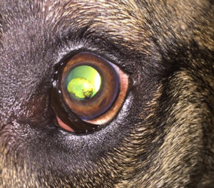
Ophthalmic Examination Findings: Iridociliary cysts are noted as unilateral or bilateral, single or multiple, circular or oval, variably sized or variably pigmented structures free-floating within the anterior chamber or attached to the pupillary margin. Pharmacologic mydriasis may reveal cysts attached within the posterior chamber. If free-floating, cysts are gravity dependent and may be seen within the ventral anterior chamber. The presence of cysts may be confirmed with transillumination as cystic structures will transmit light (versus solid mass structures).
Disease Description in this Species
Signalment
Iridociliary cysts are seen in adult dogs 7-9 years old2 with no sex-predilection reported. Golden Retrievers, Labrador Retrievers, Boston Terriers and Great Danes4 are more commonly affected.
Clinical Signs
Iridociliary cysts are noted as unilateral or bilateral, single or multiple, circular or oval, variably sized or variably pigmented structures free-floating within the anterior chamber or attached to the pupillary margin. Evidence of a previous cyst may also be seen as focal or multifocal circular, oval or irregular deposits of pigment along the corneal endothelium or anterior lens capsule. These deposits may be noted with or without concurrent iridociliary cysts.
Etiology
- Spontaneous
- Idiopathic
- Traumatic
- Congenital
- Inflammatory
Breed Predilection
- Golden Retrievers
- Labrador Retrievers
- Great Danes
- Boston Terriers
Sex Predilection
None
Age Predilection
Middle-aged (7-9yr)
Diagnostic Procedures
Iridociliary cysts are typically easily diagnosed based on appearance. Cysts are brown or black, but may rarely be tan or yellow.5 Cystic structures are differentiated from solid masses by transillumination and movement, if free-floating. High-resolution ultrasound may be used as a diagnostic aid to differentiate cysts from uveal masses if they are unable to be transilluminated or if they are very thick-walled and do not transilluminate.6 If ventral anterior chamber cysts are present, rotating the patient to dorsal recumbency will cause the cysts to float down (i.e. “snow-globe” effect), thereby delineating them from masses.
Images

A patient with a single ventral anterior chamber free-floating iridociliary cyst is depicted. Notice the shadowing effect of the cyst and transillumination. These characteristics differentiate it from an intrairidal mass.
Treatment/Management/Prognosis
Specific Therapy
Typically, uveal cysts are benign and incidental findings. They usually do not require treatment or monitoring over time. In certain breeds (see Monitoring and Prognosis), referral to a veterinary ophthalmologist for a full ophthalmic examination may be warranted for long-term monitoring and for differentiation between benign cysts and early pathologic disease. Iris cysts that are large and obstructing the pupil, causing visual disturbances, behavior changes or intraocular hypertension may be deflated using transcorneal diode laser therapy3 or manually using a 25-gauge or 27-gauge needle with magnification (i.e. operating microscope).1 Poorly pigmented cysts may not be amenable to diode laser therapy.
Supportive Therapy
None
Monitoring and Prognosis
Because most cysts are benign and incidental, the prognosis is typically excellent for most affected patients. If cysts are large, numerous or obstructing the pupillary margin, periodic monitoring for changes to vision or elevated intraocular pressure every 4-6 months may be warranted. Iridociliary cysts may be a precursor to pigmentary uveitis in Golden Retrievers,7 Great Danes8 and American Bulldogs,9 although benign cysts may also occur in these breeds, making recognition of benign versus more serious pathologic changes difficult in these breeds. In Golden Retrievers, thin-walled and attached cysts are linked to development of pigmentary uveitis while thick-walled and free-floating cysts have not been shown to progress to development of disease.10 If iridociliary cysts are diagnosed in Golden Retrievers, Great Danes or American Bulldogs, referral to a veterinary ophthalmologist for full ophthalmic evaluation should be considered.
Differential Diagnosis
- Iridal neoplasia
- Intraocular melanoma
- Pigmentary uveitis
References
- Corcoran KA, Koch SA. Uveal cysts in dogs: 28 cases (1989-1991). J Am Vet Med Assoc. 1993 Aug 15;203(4):545-6.
- Hendrix DVH. Diseases and Surgery of the Canine Anterior Uvea. In Gelatt KN (ed): Veterinary Ophthalmology 4th Pg 816 Blackwell Publishing, Ames IA
- Gemensky-Metzler AJ, Wilkie DA, Cook CS. The use of semiconductor diode laser for deflation and coagulation of anterior uveal cysts in dogs, cats and horses: a report of 20 cases. Vet Ophthalmol. 2004 Sep-Oct;7(5):360-8.
- Grahn BH1, Sandmeyer LS, Bauer BS. Diagnostic ophthalmology. Can Vet J. 2012 Mar;53(3):321-2.
- Delgado E1, Pissarra H, Sales-Luís J, Peleteiro MC. Amelanotic uveal cyst in a Yorkshire terrier dog. Vet Ophthalmol. 2010 Sep;13(5):343-7.
- Bentley E1, Miller PE, Diehl KA. Use of high-resolution ultrasound as a diagnostic tool in veterinary ophthalmology. J Am Vet Med Assoc. 2003 Dec 1;223(11):1617-22, 1599.
- Townsend WM and Gornik KR. Prevalence of uveal cysts and pigmentary uveitis in Golden Retrievers in three Midwestern states. J Am Vet Med Assoc 2013 Nov 1;243(9):1298-301.
- Spiess BM1, Bolliger JO, Guscetti F, Haessig M, Lackner PA, Ruehli MB. Multiple ciliary body cysts and secondary glaucoma in the Great Dane: a report of nine cases. Vet Ophthalmol. 1998;1(1):41-45.
- Pumphrey SA1, Pizzirani S, Pirie CG, Needle DB. Glaucoma associated with uveal cysts and goniodysgenesis in American Bulldogs: a case series. Vet Ophthalmol. 2013 Sep;16(5):377-85.
- Holly VL, Sandmeyer LS, Bauer BS, Verges L, Grahn BH. Golden retriever cystic uveal disease: a longitudinal study of iridociliary cysts, pigmentary uveitis, and pigmentary/cystic glaucoma over a decade in western Canada. Vet Ophthalmol. 2016 May;19(3):237-44.
