Published by Rachel Davis, DVM, MS, DACVO October 2017
Publication: Veterinary Information Network (VIN)
Disease Description
Intracorneal hemorrhage is a rarely described condition in older dogs seen as a variably sized focal well-demarcated area of free blood within the corneal stroma.1 Intracorneal hemorrhage is always noted with corneal vascularization; however, while corneal vascularization is a relatively common finding in dogs, intracorneal hemorrhage is rare. Intracorneal hemorrhages typically occur unilaterally with no predilection for the right versus left eye and may occasionally occur bilaterally. The most common corneal area affected is the mid-central nasal quadrant, although intracorneal hemorrhage may occur in any area.1,2 This syndrome has not been reported in cats.
Etiology
A definitive underlying etiology has not been identified; however, corneal vascularization appears to be a prerequisite for development, as would seem logical. Causes of corneal vascularization are numerous with the most common being keratoconjunctivitis sicca, corneal surgery, corneal ulceration and superficial chronic keratitis.3,4 Intracorneal hemorrhage is associated with other ocular conditions in 91% of cases, suggesting that intracorneal hemorrhage may be a secondary ocular condition.2 Additionally, 59% of dogs with intracorneal hemorrhage have underlying systemic diseases with diabetes mellitus, hyperadrenocorticism, hypothyroidism, and systemic hypertension being the most common.2 In humans, intracorneal hemorrhage has also been identified with a large variety of other ocular and systemic conditions including spontaneous formation, making identification of specific causative etiology difficult.5-8
Diagnosis
Ophthalmic Examination Findings: Intracorneal hemorrhage is noted as light to dark red, focal, well-demarcated areas of free blood within the corneal stroma typically seen at the leading edge of variably dense corneal vascularization. The most common region affected is the mid-central area of the cornea with the nasal quadrant being more commonly affected than the other quadrants, although hemorrhage may occur anywhere in the cornea.1 In most patients with intracorneal hemorrhage, other ophthalmic disease is present with keratoconjunctivitis sicca, corneal ulcers and cataracts being the most likely concurrent conditions.2 Intracorneal hemorrhage has also always been reported with corneal vascularization, thus this would be an expected concurrent finding.
Physical Examination Findings: The underlying etiology of intracorneal hemorrhage is not known; however, a recent study found that 59% of patients had underlying systemic disease.2 This finding may be indicative of a relationship between intracorneal hemorrhage and systemic disease or may be merely related to the average age of patients with intracorneal hemorrhage (11.5yr) and represent comorbidity, seen more commonly in older dogs. A thorough physical examination should be performed in all patients with intracorneal hemorrhage and baseline systemic labwork (CBC, chemistry, urinalysis) should be considered.
Disease Description in this Species
Signalment
Intracorneal hemorrhage affects older patients with the average age being 11.4yr1 in one study and 11.5yr2 in another. A wide variety of breeds have been identified with intracorneal hemorrhage. One study found no breed predisposition for this condition1 while another study found that Bichon Frises2 were overrepresented compared to the total hospital population, although this study also had a large variety of breeds affected. No sex predilection has been identified.
Clinical Signs
Intracorneal hemorrhage is noted as light to dark red, focal, well-demarcated areas of free blood within the corneal stroma typically seen at the leading edge of variably dense corneal vascularization. The most common region affected is the mid-central area of the cornea with the nasal quadrant being more commonly affected than the other quadrants. Ocular signs may also include redness or discomfort (e.g. blepharospasm, rubbing the eye/s, etc.).2 Most patients with intracorneal hemorrhage have other ophthalmic conditions present such as keratoconjunctivitis sicca, corneal ulcers or cataracts. Approximately 60% of patients affected also have systemic disease present with a large variety of conditions being identified concurrently including diabetes mellitus, hyperadrenocorticism, hypertension, hypothyroidism, degenerative heart valvular disease and many others.2 Whether the concurrent ocular conditions or systemic conditions are related to intracorneal hemorrhage or merely an indication of an aged canine population is not known.
Etiology
Unknown
Systemic disease – diabetes mellitus, hyperadrenocorticism, hypothyroidism, immune-mediated thrombocytopenia, hypertension, degenerative heart valve disease, portosystemic shunts, epilepsy, lymphoma, hyperparathyroidism, immune-mediated hemolytic anemia, Lyme disease, hemangiosarcoma, sepsis
Ocular disease – KCS, cataracts, corneal ulcers, iris atrophy, nuclear sclerosis, pseudophakia, calcium keratopathy, pigmentary keratitis, eyelid mass, glaucoma, progressive retinal atrophy, bullous retinal detachment, chronic superficial keratitis, corneal degeneration, panuveitis, sudden acquired retinal degeneration syndrome, corneal stromal abscess, asteroid hyalosis, chronic hemophthalmos, distichiasis, lens-induced uveitis, lens subluxation, nictitans gland prolapse, phthisis bulbi
Breed Predilection
None
Bichon Frise
Sex Predilection
None
Age Predilection
Older – mean age 11.4yr
Diagnostic Procedures
Diagnosis is typically readily made upon observing free blood within the corneal stroma. Routine labwork or other diagnostics may be warranted to evaluate for other systemic conditions present. Ocular diagnostics including tear evaluation, fluorescein staining and tonometry are warranted. Full slit-lamp biomicroscopy with a board-certified veterinary ophthalmologist may be warranted for diagnosis and treatment of other concurrent ocular conditions. Although not reported for use in cases of intracorneal hemorrhage to date, newer advanced corneal imaging techniques such as optic coherence tomography, biomicroscopic ultrasonography and confocal microscopy may be useful in the future to better characterize the nature and location of the hemorrhage and identify other underlying corneal pathology.9-11
Images
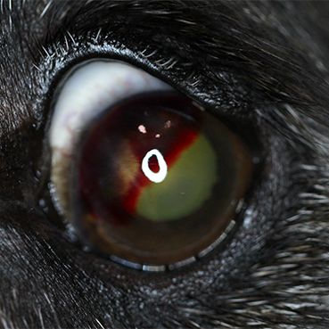
Figure 1: The left eye of a 9yr old MN Labrador mix is depicted with a large, variably dense, intracorneal hemorrhage affecting approximately 40% of the corneal surface. The hemorrhage surrounds the leading edge of admixed corneal vascularization. This patient had normal systemic labwork and was diagnosed with a qualitative tear deficiency that was treated with topical therapy.
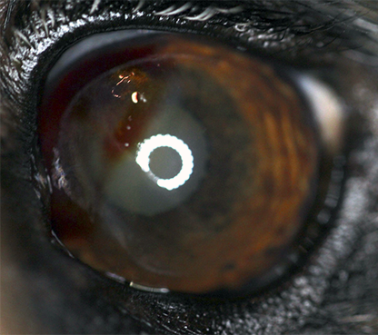
Figure 2: The same patient from Figure 1 is depicted after 4 weeks of topical therapy. The hemorrhage has significantly improved with resolution in some areas. The vascularization has regressed and the area is thinning. Yellow-brown discoloration to the corneal stroma is persistent in the area of previous hemorrhage.
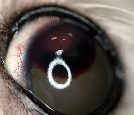
Figure 3: The left eye of a 12yr old FS Shih Tzu is depicted with a large, well-demarcated, dark red, intracorneal hemorrhage at the leading edge of a raised intracorneal stromal bed of vascularization and granulation.
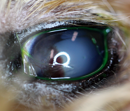
Figure 4: The left eye of a 11yr old Yorkshire terrier is depicted with a focally extensive, red, intracorneal hemorrhage at the leading edge of a large bed of corneal vascularization. This patient had concurrent blepharospasm and a qualitative tear deficiency OU that was treated with topical lacrimostimulant and anti-inflammatory therapy.
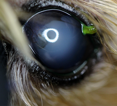
Figure 5: The same patient from Figure 4 is depicted after 4 weeks of topical therapy. The hemorrhage has resolved and marked vascular regression is noted. Mild corneal pigmentation is still present, although thinning. The patient was comfortable with resolution of the previous clinical signs. The pigment (out of focus) in the dorsonasal quadrant with an emerging strand in both photos is severe iris atrophy found as an incidental finding.
Treatment/Management/Prognosis
Specific Therapy
Specific therapy for the hemorrhage has not been identified; however, identification and treatment of other underlying ocular conditions, specifically surface disease/s, and systemic disease is warranted (see supportive therapy).
Supportive Therapy
Logically, ocular conditions that promote corneal vascularization like keratoconjunctivitis sicca and corneal ulceration3,4 are likely contributory to intracorneal hemorrhage development, although these conditions are common and intracorneal hemorrhage is rare, suggesting that there may be other unidentified factors present. Regardless, if ocular surface disease is present, treatment with lacrimostimulant (e.g. Optimmuneâ, tacrolimus) and anti-inflammatory therapy (e.g. topical steroid, topical NSAID) is warranted. Caution should be exercised when using topical steroids or NSAIDs in patients with severe keratoconjunctivitis sicca based on their risk for corneal ulceration. Lacrimostimulant therapy has also been shown to have antiangiogenic and anti-inflammatory properties, making them ideal for topical use in patients with intracorneal hemorrhage.12 Treatments reported for intracorneal hemorrhage include topical anti-inflammatories, topical antibiotics and lubricants, all with varied success.1,2
Systemic conditions should also be identified and treated, particularly those that may affect vascular integrity (e.g. diabetes mellitus, hypothyroidism) and hemodynamic health (e.g. hypertension, immune-mediated thrombocytopenia, hyperadrenocorticism).
Monitoring and Prognosis
Most patients with intracorneal hemorrhage either have stable disease with time or improvement with a variety of topical therapies.1,2 Approximately 60% of patients will have improvement over time with variable therapy (systemic and topical depending on other underlying conditions) with time to resolution being 198 days on average.2 After resolution, long term effects include yellow-brown staining in the area of previous hemorrhage, fibrosis or corneal degeneration, corneal lipidosis, pigmentation and/or stromal thinning. Persistent vascularization of the cornea was noted in approximately 30% of patients and 30% of patients did not have improvement in the intracorneal hemorrhage, although worsening was not noted. Blindness related to the intracorneal hemorrhage has not been reported.
Differential Diagnosis
Corneal neoplasia
Corneal granulation/vascularization
Hyphema
Endothelial pigment
Limbal mass
Chronic superficial keratitis
References
- Matas M, Donaldson D, Newton RJ. Intracornealhemorrhage in 19 dogs (22 eyes) from 2000 to 2010: a retrospective study. Vet Ophthalmol. 2012 Mar;15(2):86-91.
- Violette NP, Ledbetter EC. Intracornealstromal hemorrhage in dogs and its associations with ocular and systemic disease: 39 cases. Vet Ophthalmol. 2017 Jan;20(1):27-33.
- Chow DW, Westermeyer HD. Retrospective evaluation ofcorneal reconstruction using ACell Vet(™) alone in dogs and cats: 82 cases. Vet Ophthalmol. 2016 Sep;19(5):357-66.
- Gilger BC. Diseases and Surgery of the Canine Cornea and Sclera. In Gelatt KN (ed): Veterinary Ophthalmology 4th Pg 690-725. Blackwell Publishing, Ames IA
- Freiberg FJ, Parente Salgado J, Grehn F, Klink T. Intracornealhematoma after canaloplasty and clear cornea phacoemulsification: surgical management. Eur J Ophthalmol. 2012 Sep-Oct;22(5):823-5.
- Gismondi M, Brusini P. Intracorneal hematoma after canaloplasty. Eur J Ophthalmol. 2013 May-Jun;23(3):442.
- Kaiura TL, Seedor JA, Koplin RS, Rhee MK, Ritterband DC, Lipton EJ. Subepithelialintracorneal hemorrhage in a soft contact lens user. Eye Contact Lens. 2004 Jul;30(3):120-1.
- Sudha V, Burgess SE. Spontaneous intracorneal haemorrhage. Br J Ophthalmol. 1999 Jun;83(6):757-8.
- Vallone LV,Enders AM, Mohammed HO, Ledbetter EC. In vivo confocal microscopy of brachycephalic dogs with and without superficial corneal Vet Ophthalmol. 2017 Jul;20(4):294-303.
- Wolfel AE,Pederson SL,Cleymaet AM, Hess AM, Freeman KS. Canine central corneal thickness measurements via Pentacam-HR® , optical coherence tomography (Optovue iVue® ), and high-resolution ultrasound biomicroscopy. Vet Ophthalmol. 2017 Oct 15. doi: 10.1111/vop.12518. [Epub ahead of print]
- Ledbetter EC,Norman ML,Starr JK. In vivo confocal microscopy for the detection of canine fungal keratitis and monitoring of therapeutic response. Vet Ophthalmol. 2016 May;19(3):220-9.
- Bucak YY,Erdurmus M,Terzi EH, Kükner A, Çelebi S. Inhibitory effects of topical cyclosporine A 0.05% on immune-mediated corneal neovascularization in rabbits. Graefes Arch Clin Exp Ophthalmol. 2013 Nov;251(11):2555-61.
