Published by Rachel (Mathes) Davis, DVM, MS, DACVO July 2016
Publication: Veterinary Information Network (VIN)
Disease Description
Proptosis is the sudden expulsion of the globe from the orbit causing the globe to be positioned anterior to the eyelids and the eyelids to become entrapped posterior to the globe. Globe proptosis is due to orbital trauma, either blunt, shearing or penetrating. A detailed history is important to determine the type of trauma sustained, which may help the practitioner to further evaluate the patient for systemic or other craniofacial trauma. In some cases, the nature of the trauma may be inferred, but is not actually known.
Etiology
Globe proptosis occurs secondary to orbital or head trauma (e.g. dog bites, vehicular trauma). Because the feline orbit is more enclosed with bone than the canine orbit,1 severe trauma is typically sustained to displace the feline globe from the orbit.2
Diagnosis
Ophthalmic Examination Findings: Diagnosis is straightforward and made upon identification of the globe anterior to the orbit and eyelids, usually with a history of craniofacial trauma. In cats, there may be concurrent severe orbital or head trauma present often seen as periocular subcutaneous emphysema, severe soft tissue swelling, lacerations or palpable skull fractures. When assessing a proptosed globe, it is important to assess both the periocular structures and globe viability. Because cats have a shallower orbit and shorter optic nerve than dogs, traction on the optic nerve may cause contralateral blindness.3 Therefore, the contralateral eye should also be assessed for vision at the time of presentation.
Physical Examination Findings: Because globe proptosis is secondary to trauma, a thorough physical examination (+/- systemic bloodwork and imaging) should be performed to assess for any other systemic signs of trauma.
Disease Description in this Species
Signalment
Young (mean age 4.7yrs) unneutered male cats are at higher risk for globe proptosis,4 likely from an increased risk of trauma secondary to territorial, roaming and mating behavior.
Clinical Signs
Cats are typically presented on an emergent basis after sustaining head trauma. Because the appearance of a proptosed globe is somewhat disturbing aesthetically, most clients will be distraught when bringing their pet to the veterinary clinic. Diagnosis is easily made based on visual appearance of the globe anterior to the orbit and eyelids.
Etiology
Trauma
Breed Predilection
None
Sex Predilection
Intact males
Age Predilection
Young cats
Diagnostic Procedures
Ophthalmic examination – globe positioned anterior to eyelids and orbit, absent menace and palpebral reflex secondary to entrapment of eyelids posterior to globe, likely mydriasis, likely absent direct pupillary light response, likely absent indirect pupillary light response from affected to non-affected eye, +/- hyphema, +/- corneal ulceration secondary to exposure and corneal drying
Physical examination – variable periocular swelling, periorbital lacerations, subcutaneous emphysema, skull fractures, epistaxis, other systemic signs of trauma
Images

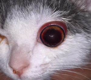
A young cat is depicted with a left globe proptosis. Notice the dark hyphema in the anterior chamber. There is also an irregular flash artifact on the corneal surface secondary to corneal drying and exposure. Also note the ipsilateral epistaxis as a result of the trauma (hit-by-car).

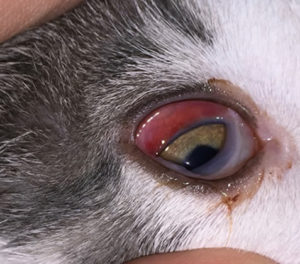
The same cat’s right eye. There is subconjunctival hemorrhage present that will resolve spontaneously over 3-5 days. This eye is visual.
Treatment/Management/Prognosis
Specific Therapy
The prognosis for vision in cats with proptosed globes is grave2,4 and other significant craniofacial injuries are often sustained in these cases. Enucleation should be considered to treat a proptosed globe in this species. Reduction of a proptosed feline globe should be viewed as a cosmetic procedure and should only be pursued if there is a high degree of certainty that the periocular and orbital structures are healthy. Because cats with craniofacial trauma often have maxillary and orbital fractures, orbital integrity, ideally with computed tomography, should be assessed prior to reduction of a feline proptosed globe.5 Enucleation should be recommended if there is significant periorbital or orbital trauma. Although globe proptosis has not been linked to feline post-traumatic ocular sarcoma, enucleation should be considered in young cats with intraocular damage concurrent with proptosis to help reduce the potential risk of this tumor later in life.6,7 Care should be taken during enucleation of a proptosed feline eye to prevent further traction on the optic nerve and subsequent contralateral blindness. Feline optic nerves should never be ligated for this reason.3 For more information on reduction of a proptosed globe, see “Globe Proptosis – Canine.”
Supportive Therapy
Non-specific systemic therapy for craniofacial and/or systemic trauma should be instituted based on the nature of the trauma and systemic state of the patient. Intravenous fluid therapy, analgesic medication and anti-inflammatory therapy should be administered if indicated.
Monitoring and Prognosis
For follow up on proptosed globe reduction surgery, see “Globe Proptosis – Canine” as the same would apply for a cat. Patients undergoing enucleation surgery are typically evaluated 10-14 days after enucleation to ensure that normal healing has occurred. Follow up for other craniofacial and systemic trauma is based on specific therapy and nature of the trauma. Recommendations to neuter an intact male cat and/or keep the cat inside may help prevent future traumatic injuries.
Differential Diagnosis
- Exophthalmos secondary to orbital mass
- Orbital fractures
References
- Samuelson, DA. Ophthalmic Anatomy. In Gelatt KN (ed): Veterinary Ophthalmology 4th Pp 38-40
- Gilger BC, et al. Traumatic ocular proptoses in dogs and cats: 84 cases (1980-1993). J Am Vet Med Assoc 1995
- Donaldson D, et al. Contralateral optic neuropathy and retinopathy associated with visual and afferent pupillomotor dysfunction following enucleation in six cats. Vet Ophthalmol 2014
- Fritsche J, et al. Prolapse of the eyeball in small animals: a retrospective study of 36 cases. Tierarztl Prax 1996
- Wunderlin N, et al. Computed tomography in cats with craniofacial trauma with regard to maxillary and orbital fractures. Tierarztl Prax 2012
- Dubielzig RR, et al. Clinical and morphologic features of post-traumatic ocular sarcomas in cats. Vet Pathol 1990
- Zeiss CJ, Johnson EM, Dubielzig RR. Feline intraocular tumors may arise from transformation of lens epithelium. Vet Pathol 2003
