Corneal Erosion, Persistent in Cats
Published by Emily A. Latham, DVM
Last updated on 5/31/2024.
Contributors:
Revised by Emily A. Latham MVB and Rachel L. Davis DVM, MS, DACVO
Revised by Pilar Camacho-Luna LV, 10/16/2019
Original author was Terah Webb DVM, DACVO, 10/10/2012
Synonyms:
Indolent corneal ulcer
Nonhealing corneal ulcer
Refractory corneal ulcer
Disease Description:
Definition
Persistent corneal erosions are uninfected, epithelial defects with a nonadherent epithelial border. Corneal stroma is intact. The lesion is characterized by failure to heal within 2 weeks via normal wound healing processes. Persistent erosions are not as common in the cat as in the dog. They can be present for weeks to months.
Anatomy
Cornea is comprised of 4 layers, namely epithelium, stroma, Descemet’s membrane, and endothelium. Epithelium contains 5-7 layers of nonkeratinized squamous, polyhedral (wing), and basal cells. Beneath the epithelium lies a basement membrane. Hemidesmosomes attach the basement membrane of the basal cells to the underlying stroma.
Superficial corneal ulcers occur with loss of the epithelial layer. Epithelial cells have great regenerative power, with a basal cellular turnover time of 7 days. Under normal circumstances, if the entire corneal epithelium is removed the cornea will resurface in most species within 48-72 hours.1 However, the basement membrane can take weeks to months to reestablish, and corneal epithelium can be easily removed until the basement membrane is reformed.34
Pathophysiology and Etiology
Pathophysiology of these ulcers in cats is not fully elucidated, however, many are thought to be caused by feline herpesvirus (FHV-1) infection.2,3,34 Cats treated with topical corticosteroids are at risk for corneal ulcers due to reactivation of latent herpesvirus. Up to 50% of clinically normal cats have FHV-1 DNA present in their corneas.4,5
Persistent erosions can also occur spontaneously or from mild, superficial trauma.
Facial conformation may play a role in the development of some ulcers because some brachycephalic breeds are predisposed.3,6 It is speculated that the combination of prominent eyes; low sub-epithelial and sub-basal corneal nerve fiber density; and lower central corneal sensitivity may make brachycephalic cats more susceptible to chronic ulcerative keratitis.7,8 Eyelid abnormalities (e.g. entropion) are also more common in brachycephalic cats and can irritate the cornea.34
Diagnosis
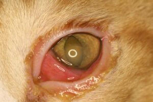
Physical Examination Findings/History: History of a chronic (>2 weeks), nonhealing corneal ulcer is a characteristic feature.2 Pain is variable. Varying degrees of epiphora, blepharospasm, and conjunctival hyperemia may be present (Figure 1A).
Ocular Examination Findings: The ulcer can occur in any quadrant of the cornea and is very superficial. In cats with FHV-1 infection, ulcers may start as punctate, linear, or branching (dendritic) lesions and progress to geographic, larger ulcers. A ring of loose epithelium surrounds the erosion and creates a halo when retroilluminated (Figure 2). Fluorescein diffuses under the loose epithelium (Figure 3). If loose epithelium is not obvious and the ulcer is superficial, a topical anesthetic agent (e.g. proparacaine) can be applied and the ulcer site gently rubbed with a sterile cotton swab (Figure 4). If epithelium moves with the swab, diagnosis is confirmed.
Superficial corneal vascularization can occur but is variable. Degree of vascularization partially depends on size and duration (chronicity) of the ulcer (Figure 5). Development of granulation tissue with chronic erosions is less common than in dogs. Focal stromal edema may be present under the ulcer (Figure 1A). The presence of diffuse corneal edema is suggestive of endothelial disease and may represent a contributing cause of the erosion. Persistent corneal erosions do not cause diffuse corneal edema. Most persistent erosions are unilateral but bilateral ulcers can occur. Erosions may also occur in the contralateral eye at some other time in the cat’s life. Most eyes have only one erosion; however, multiple sites can be affected.
Secondary uveitis, stromal infection (e.g. yellow, white discoloration), and stromal malacia are not components of persistent erosions. With the exception of FHV-1, other primary causes of corneal ulcers and delayed wound healing are absent.
Cytology: Cytology is not required to make a diagnosis. However, if corneal cytology is performed it often shows mild, neutrophilic inflammation.2 Infectious organisms are absent.
Culture: Culturing of the cornea is not usually necessary and does not yield growth of microorganisms.
Other Tests: For information on diagnosing FHV-1 infection, see the Feline VINcyclopedia chapter on FHV-1 Ocular Infection.
Disease Description in This Species:
Signalment
In two studies, the Persian, Burmese, and exotic shorthair were overrepresented, possibly indicating a predisposition for brachycephalic breeds.3,6,34 Although erosions can occur at any age, cats are often younger than affected dogs because of FHV-1 infections. No sex predisposition has been reported.
Clinical Signs
Potential signs include tearing, tear staining near the medial canthus, blepharospasm, pawing or rubbing at the eye, conjunctival redness, and visible cloudiness in the cornea. With FHV-1 involvement, stromal keratitis can develop with chronic lesions, and other signs may be present, such as lethargy, anorexia, fever, sneezing, coughing, and nasal discharge.
Cats with ulcers that fail to heal for several weeks are at high risk for developing a sequestrum within the exposed corneal stroma.11 Occasionally, entropion develops secondary to ocular pain associated with corneal ulcers or conjunctivitis. Entropion exacerbates the pain response as hairs contact the cornea, and secondary irritation may cause or hasten the development of corneal sequestra.
Etiology:
Feline herpesvirus-1 (FHV-1)
Idiopathic, unknown
Trauma
Breed / Species Predilection:
Burmese
Exotic shorthair
Persian
Sex Predilection:
None
Age Predilection:
Young adult
Clinical Findings:
AFEBRILE
Anorexia, hyporexia
Blepharospasm, eye pain
Conjunctival congestion, hyperemia
CONJUNCTIVITIS
Corneal opacity
Corneal ulcer, keratitis
Corneal vascularization
Depression, lethargy
Edema corneal
EDEMA or SWELLING
Epiphora, lacrimation increased
Eyelid pruritus, rubbing
Keratoconjunctivitis
NASAL DISCHARGE
Nasal discharge mucopurulent
Nasal discharge purulent
Nasal discharge serous
OCULAR DISCHARGE
Ocular discharge mucoid
Ocular discharge purulent
Ocular discharge serous
PAIN
Third eyelid, nictitating membrane prolapsed
ULCERS
ZZZ INDEX ZZZ
| Diagnostic Procedures: | Diagnostic Results: | |
| Cytology, biopsy of conjunctiva or cornea | Lymphocytic infiltration | |
| Neutrophilic infiltration of conjunctiva | ||
| Ocular examination | Corneal dendritic ulcers | |
| Corneal fluorescein staining positive | ||
| Corneal geographic ulcer | ||
| Loose epithelium over corneal erosion | ||
| PCR assay | Herpesvirus-1 detected by PCR |
Images:

2-yr-old cat with worsening conjunctival redness and ocular discharge over several weeks. The hazy area in the cornea behind the ring flash is an axial superficial erosion. Clinical findings were compatible with herpesvirus infection.

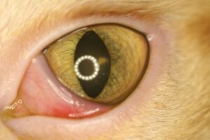
2-yr-old cat. Photo taken 3 weeks after starting anti-viral medication. Corneal ulcer has healed and conjunctival hyperemia has improved. Small area of fibrosis is visible adjacent to the ring flash at 2 o’clock.
Treatment / Management:
SPECIFIC THERAPY
Specific therapy involves removal of all loose epithelium, with administration of antiviral agents if FHV-1 is suspected as an etiology.
Debridement
Under a topical anesthetic (e.g. proparacaine) debridement is performed using a sterile cotton swab. As the swab is rubbed from the center of the ulcer towards the edge, it catches, lifts, and strips off loose epithelium. Normal, adherent epithelium cannot be removed with a cotton swab, so debridement should extend peripherally until no further epithelium can be removed. Adequate time must be given for new epithelium to migrate across the defect and adhere to the underlying stroma, so debridement should not be performed more frequently than every 10-14 days.
Debridement and Grid/Punctate Keratotomy
Grid or punctate keratotomy are not performed in cats because of the risk of corneal sequestrum formation following the procedure.6
Diamond Burr Debridement
In one study, 17/21 eyes (81%) healed with diamond burr debridement followed by insertion of a bandage contact lens.35 Recurrence rate was 17.6% and corneal sequestrum formation occurred in one eye.35 Burmese cats were overrepresented in the cases that failed to heal or experienced recurrence.35 The procedure carries the risk of sequestrum formation and has a slightly lower success rate than the superficial keratectomy.35
Superficial Keratectomy
Superficial keratectomy is an effective treatment for erosions refractory to medical treatment, with a success rate of about 85% within 4 weeks after surgery.3 It is speculated that the main advantage of superficial keratectomy is physical removal of virus, associated stromal inflammatory infiltrates, and necrotic tissue that may be impairing healing.12
SUPPORTIVE THERAPY
Appropriate therapy should be administered for FHV-1 ocular infection and associated adnexal disease.34 An Elizabethan collar is often applied to prevent self-trauma that can worsen the ulcer. Keep in mind that corneal pain is often transiently worse after debridement and the above procedures. Topical antibiotics are administered q 8-12 hrs to prevent secondary infection. Topical oxytetracycline has been shown to promote mesenchymal transformation of epithelial cells and encourage faster healing.
Analgesics are indicated because erosions can cause pain from exposure of corneal nerves and from reflex ciliary spasm. Atropine 1% or morphine 1% may be administered topically. Prolonged use of atropine can decrease tear production. Ointment preparations are preferred over solutions as they induce less salivation in cats. Systemic analgesics (gabapentin 5-10 mg/kg PO q 12-24 hrs; buprenorphine 0.005-0.01 mg/kg IM, SC; tramadol 2-4 mg/kg PO q 12 hrs) and nonsteroidal anti-inflammatory drugs may be considered.21,23 Topical proparacaine is not indicated for chronic pain control and should not be used on a repeated basis. The drug is toxic to corneal epithelium and exhibits tachyphylaxis, i.e. it becomes less effective with recurrent application.
Bandage contact lens may decrease time to healing. Contact lenses also alleviate pain associated with the eyelids rubbing against the corneal erosion.3,33
Various topical medications have been tried that are designed to promote healing by improving the bond between epithelium and stroma; by decreasing mechanical disruption of epithelial movement/adhesion from eyelid movement (i.e. blinking); or by lubricating the cornea. They include ophthalmic chondroitin sulfate solution (not available in the USA), hyaluronic acid gel, polysulfated glycosaminoglycans, and topical epidermal growth factor.22-26 For persistent erosions not associated with FHV-1, the use of heterologous serum or platelet-rich plasma does not improve healing rates.27,28 However, some evidence exists that administration of topical serum may be helpful for herpetic keratitis unresponsive to conventional treatments, particularly in cats with recent ulcers.29
MONITORING and PROGNOSIS
Ulcers are typically rechecked every 10-14 days; however, visits may be scheduled more frequently if clinical signs worsen. Persistent erosions can be frustrating to treat and often require prolonged therapy. Anti-anxiety medications (e.g. gabapentin, trazadone) may help decrease the stress of recheck visits, especially if FHV-1 is thought to be a contributing factor. The risk of progression of a chronic, nonhealing ulcer to a sequestrum should be discussed with the owner early in the course of treatment.30-32
Preventive Measures:
Use of topical, ophthalmic steroids should be reserved for feline corneoconjunctival diseases that specifically require them (e.g. eosinophilic keratoconjunctivitis) in order to limit the risk of FHV-1 activation or recrudescence.
Special Considerations:
Other Resources
Recent VIN Message board discussions on persistent corneal erosions
Proceedings articles that discuss nonhealing ulcers
Client handout on corneal ulcers and erosions in dogs and cats
2014 VIN Rounds on nonhealing corneal ulcers/erosions
Ophthalmology Fun Case 130
VIN Mentor Procedures video on corneal debridement, diamond burr keratotomy, grid keratotomy, and contact lens placement
For more images, see the Corneal Ulcers & Wounds – Cat slideshow in the Image Library
Differential Diagnosis:
Corneal sequestrum
Other causes of corneal vascularization
Other types of ulcerative keratitis
References:
- Ledbetter EC, Gilger BC: Diseases of the Canine Cornea and Sclera. In: Gelatt KN (ed): Veterinary Ophthalmology, 5th ed. Wiley-Blackwell, Ames IA 2013 pp. 976.
- Cullen CL, Wadowska DW, Singh A, et al: Ultrastructural findings in feline corneal sequestra. Vet Ophthalmol 2005 Vol 8 (5) pp. 295-303.
- Jégou J, Tromeur F: Superficial keratectomy for chronic corneal ulcers refractory to medical treatment in 36 cats. Vet Ophthalmol 2015 Vol 18 (4) pp. 335-40.
- Townsend WM, Stiles J, Guptill-Yoran L, et al: Development of a reverse transcriptase-polymerase chain reaction assay to detect feline herpesvirus-1 latency associated transcripts in the trigeminal ganglia and corneas of cats that did not have clinical signs of ocular disease. Am J Vet Res 2004 Vol 65 (3) pp. 314-19.
- Stiles J, McDermott M, Bigsby D, et al: Use or nested polymerase chain reaction to identify feline herpesvirus in ocular tissue from clinically normal cats and cats with corneal sequestra or conjunctivitis. Am J Vet Res 1997 Vol 58 (4) pp. 338-42.
- LaCroix NC, van der Woerdt A, Olivero DK: Nonhealing corneal ulcers in cats: 29 cases (1991-1999). Am Vet Med Assoc 2001 Vol 218 (5) pp. 733-35.
- Blocker T, Van Der Woerdt A: A comparison of corneal sensitivity between brachycephalic and Domestic Short-haired cats. Vet Ophthalmol 2001 Vol 4 (2) pp. 127-30.
- Kafarnik C, Fritsche J, Reese S: Corneal innervation in mesocephalic and brachycephalic dogs and cats: assessment using in vivo confocal microscopy. Veterinary Ophthalmology 2008 Vol 11 (6) pp. 363-67.
- Bentley E, Abrams GA, Covitz D, et al: Morphology and immunohistochemistry of spontaneous chronic corneal epithelial defects (SCCED) in dogs. Invest Ophthalmol Vis Sci 2001 Vol 42 (10) pp. 2262-69.
- Murphy CJ, Marfurt CF, McDermott A, et al: Spontaneous chronic corneal epithelial defects (SCCED) in dogs: Clinical features, innervation, and effect of topical SP, with or without IGF-1. Invest Ophthalmol Vis Sci 2001 Vol 42 (10) pp. 2252-61.
- Stiles J: Feline Ophthalmology. In: Gelatt KN, Gilger BC, Kern TJ (eds): Veterinary Ophthalmology, 5th ed. Wiley-Blackwell, Ames IA 2013 pp. 1477-1559.
- Deshpande SP, Zheng M, Lee S, et al: Mechanisms of pathogenesis in herpetic immunoinflammatory ocular lesions. Vet Microbiol 2002 Vol 86 (1-2) pp. 17-26.
- Thomasy SM, Maggs DJ: A review of antiviral drugs and other compounds with activity against feline herpesvirus type 1. Vet Ophthalmol 2016 Vol 19 (suppl 1(0)) pp. 119-30.
- Romanowski EG, Bartels SP, Gordon YJ: Comparative antiviral efficacies of cidofovir, trifluidine, and acyclovir in the HSV-1 rabbit keratitis model. Invest Ophthalmol Vis Sci 1999 Vol 40 (2) pp. 378-84.
- Sebbag L, Thomasy SM, Woodward AP, et al: Pharmacokinetic modeling of penciclovir and BRL42359 in the plasma and tears of healthy cats to optimize dosage recommendations for oral administration of famciclovir. Am J Vet Res 2016 Vol 77 (8) pp. 833-45.
- Maggs DJ, Nasisse MP, Kass PH: Efficacy of oral supplementation with L-lysine in cats latently infected with feline herpesvirus. Am J Vet Res 2003 Vol 64 (1) pp. 37-42.
- Bol S, Bunnik EM: Lysine supplementation is not effective for the prevention or treatment of feline herpesvirus 1 infection in cats: a systematic review. BMC Vet Res 2015 Vol 11 (0) pp. 284.
- Sandmeyer LS, Keller CB, Bienzle D: Effects of interferon-alpha on cytopathic changes and titers for feline herpesvirus-1 in primary cultures of feline corneal epithelial cells. Am J Vet Res 2005 Vol 66 (2) pp. 210-6.
- Slack JM, Stiles J, Leutenegger CM, et al: Effects of topical ocular administration of high doses of human recombinant interferon alpha-2b and feline recombinant interferon omega on naturally occurring viral keratoconjunctivitis in cats. Am J Vet Res 2013 Vol 74 (2) pp. 281-89.
- Spatola RA, Thangavelu M, Chandler HL: The effects of topical nalbuphine on canine corneal cells in vitro. In: Abstract presented at the Annual Conference of The American College (ed): Proceed Am Coll Vet Ophthalmol 2009 Vol 12 (6) pp. 390-409.
- Clark JS, Bentley E, Smith LJ: Evaluation of topical nalbuphine or oral tramadol as analgesics for corneal pain in dogs: a pilot study. Vet Ophthalmol 2011 Vol 14 (6) pp. 358-64.
- Ledbetter RJ, Munger RJ, Ring RD, et al: Efficacy of two chondroitin sulfate ophthalmic solutions in the therapy of spontaneous chronic corneal epithelial defects and ulcerative keratitis associated with bullous keratopathy in dogs. Vet Ophthalmol 2006 Vol 9 (2) pp. 77-87.
- Miller WW: Using polysulfated glycosaminoglycan to treat persistent corneal erosions in dogs. Vet Med 1996 Vol 91 (12) pp. 916-22.
- Yang G, Espandar L, Mamalis N, et al: A cross-linked hyaluronan gel accelerates healing of corneal epithelial abrasion and alkali burn injuries in rabbits. Vet Ophthalmol 2010 Vol 13 (3) pp. 144-50.
- Williams DL, Wirostko BM, Gum G, et al: Topical Cross-Linked HA-Based Hydrogel Accelerates Closure of Corneal Epithelial Defects and Repair of Stromal Ulceration in Companion Animals. Invest Ophthalmol Vis Sci 2017 Vol 58 (11) pp. 4616-22.
- Kirschner SE, Brazzell RK, Stern ME, et al: The use of topical epidermal growth factor for treatment of nonhealing corneal erosions in dogs. J Am Anim Hosp Assoc 1991 Vol 27 (4) pp. 449-52.
- Eaton JS, Hollingsworth SR, Holmberg BJ, et al: Effects of topically applied heterologous serum on reepithelialization rate of superficial chronic corneal epithelial defects in dogs. J Am Vet Med Assoc 2017 Vol 250 (9) pp. 1014-22.
- Edelmann ML, Mohammed HO, Wakshlag JJ, et al: Clinical trial of adjunctive autologous platelet-rich plasma treatment following diamond-burr debridement for spontaneous chronic corneal epithelial defects in dogs. J Am Vet Med Assoc 2018 Vol 253 (8) pp. 1012-21.
- Kim SE, Lee M, Seo K: Clinical application of serum eye drops for herpetic keratitis in cats: a pilot study. Int J App Res Vet Med 2018 Vol 16 (3) pp. 221-25.
- Morgan RV: Feline corneal sequestration: A retrospective study of 42 cases (1987-1991). J Am Anim Hosp Assoc 1994 Vol 30 (1) pp. 24-28.
- Startup FG: Corneal necrosis and sequestration in the cat: A review and record of 100 cases. J Small Anim Pract 1988 Vol 29 (7) pp. 476-86.
- Featherstone HJ, Sansom J: Feline corneal sequestra: A review of 64 cases (80 eyes) from 1993-2000. Vet Ophthalmol 2004 Vol 7 (4) pp. 213-27.
- Morgan R, Abrams K: A comparison of six different therapies for persistent corneal erosions in dogs and cats. Vet Comp Ophthalmol 1994 Vol 4 (1) pp. 38-43.
- Glaze MB, Maggs DJ, Plummer CE: Feline ophthalmology. In: Gelatt KN, Ben-Schlomo G, Gilberg BC, et al (eds): Veterinary Ophthalmology, 6th ed. John Wiley & Sons, Hoboken NJ 2021 pp. 1645-1880.
- Anastassiadis Z, Bayley KD, Read RA : Corneal diamond burr debridement for superficial non-healing corneal ulcers in cats. 2022 Vol 25 (6) pp. 376-82.
Feedback:
If you note any error or omission or if you know of any new information, please send your feedback to VINcyclopedia@vin.com.
If you have any questions about a specific case or about this disease, please post your inquiry to the appropriate message boards on VIN.
