Publication: Veterinary Information Network (VIN)
Disease Description
Cataract (or cataracts) refers to any opacification of the intraocular crystalline lens.1 The lens is composed of an external capsule made mostly of collagen and internal precisely organized lens fibers made of approximately 60% protein and 40% water. While the biomechanical and physiologic processes of the lens are very complex, the lens is composed of basically one substance, protein. Thus, the lens’s response to a variety of insults results in essentially one basic outcome, that of cataract formation. Cataract formation is, therefore, not specific to any underlying pathologic process, but merely denotes an abnormality of the lens.
Cataracts are typically characterized based on overall involvement of the lens and severity. Incipient cataracts affect <15% of the lens (figures), immature cataracts involve 15-100% of the lens and are not dense enough to abolish the tapetal reflection (figure), mature cataracts involve the entire lens and prevent the tapetal reflection and hypermature cataracts involve the entire lens and have advanced to resorption seen as hyperreflective, “sparkly” area within the lens.1 Hypermature cataracts often have associated lens wrinkling and significant lens-induced uveitis.2
Etiology
Cataracts may be caused by a variety of underlying pathologic processes including nutrition,3,4 genetic factors,5 endocrine disease,6 trauma,7-10 metabolic disorders,11,12 intraocular inflammation,13 external or internal toxins,14 progressive retinal atrophy15 and age-related disease.15 The most common cause of cataracts in purebred dogs is inherited disease5,15,16 with over 150 canine breeds being shown or suspected to be affected with inherited cataracts.1 The second leading cause of cataracts in dogs is diabetes mellitus.6,17,18
Canine diabetes mellitus causes cataract formation in this species due to relatively high levels of aldose reductase, an enzyme that promotes conversion of glucose to sorbitol.19,20 Sorbitol, a large, hyperosmotic molecule, concentrates in the lens and causes diffusion of water into the lens in the diabetic state due to high levels of serum glucose. The increased water imbibition into the lens causes disruption of lens fibers and cataract formation. Cats and humans have relatively low levels of aldose reductase which is why cataract formation secondary to diabetes mellitus is uncommon in these species.17,18 Canine patients with diabetic cataracts have also been shown to have higher levels of aqueous humor fibrinolytic activity,21 but not vascular endothelial growth factor,22 than nondiabetic dogs. The significance of these findings require further investigation, but may suggest differences in underlying pathophysiology of diabetic versus nondiabetic cataract formation.
Diagnosis
Ophthalmic Examination Findings: Cataracts are seen as an opacification of the lens. This may be observed easily with retroillumination of the eye after pharmacologic pupil dilation (i.e. tropicamide or atropine administration topically). Retroillumination is performed by aiming a focal light source into the eye at arm’s distance away and at the observer’s eye level. Small opacifications of the lens may be observed as dark or light areas within the lens (i.e. areas of opacification that are impeding passage of light back) (figure). Retroillumination will also allow the observer to evaluate for a tapetal reflection (or fundic reflection in atapetal dogs) (figure), thereby allowing differentiation between immature and mature cataracts if the entire lens is affected. Direct focal illumination with the light source close to the eye and the observer in front of the eye may also be performed and incipient or immature cataracts will appear as “white” areas within the lens or, if the entire lens is involved, the whole lens to appear as white. (figures)
Ophthalmic evaluation with retroillumination and direct focal illumination will help differentiate cataracts from nuclear sclerosis, a normal aging change of the lens caused by increased density of nuclear lens fibers. The increased lens fiber density imparts a gray or milky color to the lens. Unlike cataracts, nuclear sclerosis does not cause impedance of light passage through the lens. Thus, the tapetal reflection will not be altered nor will the lens appear as white using retroillumination and direct focal illumination respectively.
Identification of the location of the cataract within the lens and further characterization of the cataracts typically require advanced training and slit-lamp biomicroscopy performed by a veterinary ophthalmologist.
Physical Examination Findings: Because some causes of canine cataracts are related to systemic (e.g. diabetes mellitus) or other ocular disease (e.g. trauma, uveitis), secondary factors contributing to cataract formation should be identified. A full physical examination, clinical history and, when warranted, a comprehensive hematologic and blood chemistry, should be pursued for patients presenting with cataracts.
Disease Description in this Species
Signalment
- Any breed, sex or age of dog may be affected by cataracts.
- Inherited cataracts are more common in young to middle-aged purebred dogs.1,16
- Diabetic cataracts are more common in middle-aged, female dogs.6
Clinical Signs
White static or progressive opacification of the lens. See ophthalmic examination.
Sex Predilection
Any
Age Predilection
Young to middle-aged
Diagnostic Procedures
Ophthalmic examination: See ophthalmic examination
Slit-lamp biomicroscopy: Full ophthalmic evaluation using slit-lamp biomicroscopy with a veterinary ophthalmologist is warranted for some patients affected by cataracts, especially if the owner is pursuing or interested in cataract surgery or in breeding animals.
Ocular ultrasound: Evaluation for cataracts is best performed by using multiple types of illumination during ophthalmic examination with pharmacologic mydriasis; however, ocular ultrasound may be used to evaluate for cataracts or lens integrity if anterior ocular pathology precludes evaluation of the lens partially or entirely.8,20 Cataracts are seen ultrasonographically as hyperechoic uniform or irregular opacification within the lens. High-resolution ocular ultrasound may also be used to identify lens capsular ruptures or tears prior to cataract surgery or as a cause of fulminate uveitis.
Orthopedic Foundation of America (OFA) Ophthalmic Certification Examination: Examination by a board-certified veterinary ophthalmologist is warranted for any purebred dogs being used for breeding purposes. Selective breeding has been shown to decrease the incidence of cataracts in breeding populations.23 In the United States, the OFA oversees and certifies evaluation of breeding animals.
Systemic Evaluation: A full physical examination, clinical history and, when warranted, a comprehensive hematologic and blood chemistry, should be pursued for patients presenting with cataracts.
Images
Figure 1A,B, C and D: A patient with incipient cataracts is shown using retroillumination and focal direct illumination with pharmacologic mydriasis OS and OD. Note that the cataract appears as a yellowish opacity that alters the tapetal reflection using retroillumination while it appears as a white opacification using focal direct illumination.
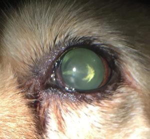
Figure 1A – Cataract – Canine
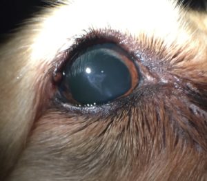
Figure 1B – Cataract – Canine
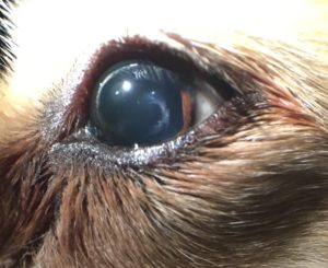
Figure 1C – Cataract – Canine
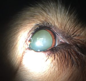
Figure 1D – Cataract – Canine
Figure 2: An immature cataract is shown using retroillumination. The cataract affects the entire lens, but does not completely abolish the tapetal reflection, seen as a yellow reflection through the cataract. Parts of the cataract block the tapetal reflection, indicating that this cataract is moving to a mature cataract.
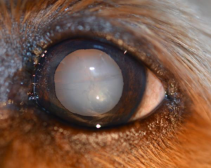
Figure 2 – Cataract – Canine
Figure 3: A mature cataract is depicted focal direct illumination. The cataract appears affects the entire lens and appears as a white structure using either retroillumination or focal direct illumination. The tapetal reflection is abolished. There is a ring-flash artifact in the center of the photograph.
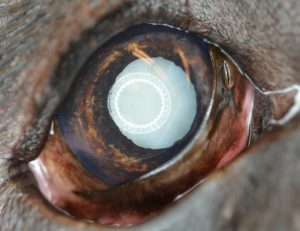
Figure 3 – Cataract – Canine
Figure 4: A hypermature cataract is depicted with resorption and irregular sparkly or hyperreflective areas. This lens also has a brown to yellow discoloration secondary to severe lens-induced uveitis. There is hemorrhage along the pupillary margin along with dyscoria and iris discoloration secondary to chronic inflammation.
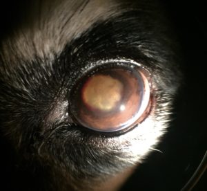
Figure 4 – Cataract – Canine
Figure 5A and B: An artificial intraocular lens implant is depicted in two patients with pharmacologic mydriasis immediately after cataract surgery.
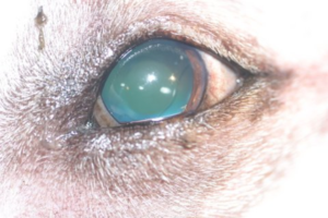
Figure 5A – Cataract – Canine
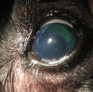
Figure 5B – Cataract – Canine
Treatment/Management/Prognosis
Specific Therapy
The standard-of-care for complete cataracts causing vision loss is lens removal using phacoemulsification with intraocular artificial lens implantation (figure).24-27 This surgery is performed by a veterinary ophthalmologist and carries a very high success rate (80-95%).24-27 Bilateral surgery is recommended when both eyes are candidates for surgery as sequential bilateral surgery carries the same success rate as surgery performed on the eyes individually at separate times.28 The success of cataract surgery is affected by breed of dog, cataract stage, level of lens-induced uveitis, duration of the cataract, lens stability (i.e. presence of lens luxation or subluxation) and other intraocular disease.24-28 It is thus important to promptly refer canine patients affected with large or complete cataracts to an ophthalmologist for further evaluation. These patients should be assessed and cataract surgery discussed if the clients are interested in pursuing vision-saving treatment. Diabetes, even if not tightly controlled, does not affect cataract surgery success and cataract surgery may be pursued early after cataract formation in these patients.25,29 In addition, the level of hyperglycemia (i.e. control of diabetes) has not been shown to be correlated to cataract formation.17 While diabetic control is considered important for other reasons, it does not prevent cataract formation.
Cataract surgery may still be an option for therapy even for patients with advanced or chronic cataracts. Referral should be considered for any patient affected with cataracts. Cataracts associated with large lens capsular tears or capsular tears caused by intumescent (swollen) cataracts are considered a surgical emergency and should be urgently referred for surgery (see phacoclastic uveitis).30
Recently, topical aldose reductase inhibitors (e.g. Kinostatã) have been shown to delay and/or prevent cataract formation in diabetic dogs.31 These medications are not yet commercially available and are costly. In addition, cessation or interruption of topical therapy at BID-TID dosing, even if only for a short time (1-3 days), has been shown to cause rapid formation of complete cataracts.32 For these reasons, topical aldose reductase inhibitor therapy has not become a popular or reasonable therapy to prevent diabetic cataracts, but may become a treatment option in the future.
No other medical therapy, to date, has been shown to delay or prevent cataract progression or formation. As in human medicine, cataracts remain a surgical disease in veterinary medicine. This is important to relay to clients, especially with the pervasiveness of false and inaccurate internet information (e.g. “dr. google”).
Supportive Therapy
In patients for which cataract surgery is not an option (e.g. severe cardiac disease precluding anesthesia, clients have declined surgery, etc.), supportive topical therapy should be instituted long-term to help prevent complication such as lens-induced uveitis and glaucoma secondary to cataracts.2,33 Topical steroidal medication (e.g. dexamethasone, prednisolone acetate) and topical non-steroidal medication (e.g. flurbiprofen, diclofenac, ketorolac) may be used BID-TID together or individually to treat lens-induced uveitis. This is important because, as cataracts progress, the incidence of lens-induced uveitis increases. Topical anti-inflammatory therapy may also be instituted prior to referral to an ophthalmologist, even if the clients are planning on pursuing cataract surgery, as some level of lens-induced uveitis is present with most cataracts. Prior to institution of long-term topical steroid therapy, fluorescein staining and evaluation of the pre-ocular tear film should be performed as steroids may worsen corneal ulceration or promote corneal malacia.
Monitoring and Prognosis
Because cataracts may cause vision loss or blindness, client education regarding monitoring for progression of incipient or small immature cataracts is warranted. In addition, because the incidence of cataract formation in diabetic dogs his very high, client education concerning likely cataract formation in newly diagnosed diabetic dogs is important. Referral to a veterinary ophthalmologist should be considered for patients affected by cataracts, especially if the clients are interested in pursuing cataract surgery for blinding cataracts.
If cataract surgery is not an option for particular patients, long term anti-inflammatory therapy (see supportive therapy) and evaluation of intraocular pressure Q4-6mo is warranted to monitor for other painful ocular conditions. Glaucoma secondary to advanced cataracts tends to be poorly responsive to topical anti-tensive therapy, thus enucleation should be considered for blind, painful globes with advanced disease.
Differential Diagnosis
- Nuclear sclerosis
- Lens luxation
References
- Davidson MG and Nelms SR. Diseases of the Canine Lens and Cataract Formation. In Gelatt KN (ed): Veterinary Ophthalmology 4th Pg 859-887. Blackwell Publishing, Ames IA.
- van der Woerdt A, Nasisse MP, Davidson MG. Lens-induced uveitis in dogs: 151 cases (1985-1990). J Am Vet Med Assoc. 1992 Sep 15;201(6):921-6.
- Quam, D. D., Morris, J. G. & Rogers, Q. R. (1987) Histidine requirement of kittens for growth, haematopoiesis and prevention of cataracts. J. Nutr. 58:521-532.
- Martin, C. L. & Chambreau, T. (1982) Cataract production in experimentally orphaned puppies fed a commercial replacement for bitch’s milk. Am. Anim. Hosp. Assoc. 18:115-119.
- Bellumori TP, Famula TR, Bannasch DL, et al. Prevalence of inherited disorders among mixed-breed and purebred dogs: 27,254 cases (1995-2010). J Am Vet Med Assoc. 2013 Jun 1;242(11):1549-55.
- Beam S, Correa MT, Davidson MG. A retrospective-cohort study on the development of cataracts in dogs with diabetes mellitus: 200 cases. Vet Ophthalmol. 1999;2(3):169-172.
- Bell CM, Pot SA, Dubielzig RR. Septic implantation syndrome in dogs and cats: a distinct pattern of endophthalmitis with lenticular abscess. Vet Ophthalmol. 2013 May;16(3):180-5.
- Paulsen ME, Kass PH. Traumatic corneal laceration with associated lens capsule disruption: a retrospective study of 77 clinical cases from 1999 to 2009. Vet Ophthalmol. 2012 Nov;15(6):355-68.
- Tetas Pont R, Matas Riera M, Newton R, Donaldson D. Corneal and anterior segment foreign body trauma in dogs: a review of 218 cases. Vet Ophthalmol. 2016 Sep;19(5):386-97.
- Martins BC, Plummer CE, Gelatt KN, et al. Ophthalmic abnormalities secondary to periocular or ocular snakebite (pit vipers) in dogs–11 cases (2012-2014). Vet Ophthalmol. 2016 Mar;19(2):149-60.
- Russell NJ, Bond KA, Robertson ID,et al. Primary hypoparathyroidism in dogs: a retrospective study of 17 cases. Aust Vet J. 2006 Aug;84(8):285-90.
- Bruyette DS, Feldman EC. Primary hypoparathyroidism in the dog. Report of 15 cases and review of 13 previously reported cases. J Vet Intern Med. 1988 Jan-Mar;2(1):7-14.
- Wooff PJ, Dees DD, Teixeria L. Aspergillus spp. panophthalmitis with intralenticular invasion in dogs: report of two cases. Vet Ophthalmol. 2016 Sep.
- Schiavo DM, Field WE, Vymetal FJ. Cataracts in beagle dogs given diazoxide. 1975 Dec;24(12):1041-9.
- Donzel E, Arti L, Chahory S. Epidemiology and clinical presentation of canine cataracts in France: a retrospective study of 404 cases. Vet Ophthalmol. 2016 Apr 7.
- Baumworcel N, Soares AM, Helms G, et al. Three hundred and three dogs with cataracts seen in Rio de Janeiro, Brazil. Vet Ophthalmol. 2009 Sep-Oct;12(5):299-301.
- Salgado D, Reusch C, Spiess B. Diabetic cataracts: different incidence between dogs and cats. Schweiz Arch Tierheilkd. 2000 Jun;142(6):349-53.
- Wilkie DA, Gemensky-Metzler AJ, Colitz CM, Bras ID, Kuonen VJ, Norris KN, Basham CR. Canine cataracts, diabetes mellitus and spontaneous lens capsule rupture: a retrospective study of 18 dogs. Vet Ophthalmol. 2006 Sep-Oct;9(5):328-34.
- Richter M, Guscetti F, Spiess B. Aldose reductase activity and glucose-related opacities in incubated lenses from dogs and cats. Am J Vet Res. 2002 Nov;63(11):1591-7.
- Salgado D1, Forrer RS, Spiess BM. Activities of NADPH-dependent reductases and sorbitol dehydrogenase in canine and feline lenses. Am J Vet Res. 2000 Oct;61(10):1322-4.
- Escanilla N, Leiva M, Monreal L, et al. Aqueous humor fibrinolytic activity in dogs with cataracts. Vet Ophthalmol. 2013 Nov;16(6):409-15.
- Abrams KL, Stabila PF, Kauper K, et al. Vascular endothelial growth factor in diabetic and nondiabetic canine cataract patients. Vet Ophthalmol. 2011 Mar;14(2):93-9.
- Koll S, Reese S, Medugorac I, Rosenhagen CU, Sanchez RF, Köstlin R. The effect of repeated eye examinations and breeding advice on the prevalence and incidence of cataracts and progressive retinal atrophy in German dachshunds over a 13-year period. Vet Ophthalmol. 2016 Apr 13.
- Klein HE, Krohne SG, Moore GE. Postoperative complications and visual outcomes of phacoemulsification in 103 dogs (179 eyes): 2006-2008. Vet Ophthalmol. 2011 Mar;14(2):114-20.
- Sigle KJ, Nasisse MP. Long-term complications after phacoemulsification for cataract removal in dogs: 172 cases (1995-2002). J Am Vet Med Assoc. 2006 Jan 1;228(1):74-9.
- Johnstone N, Ward DA. The incidence of posterior capsule disruption during phacoemulsification and associated postoperative complication rates in dogs: 244 eyes (1995-2002). Vet Ophthalmol. 2005 Jan-Feb;8(1):47-50.
- Lim CC, Bakker SC, Waldner CL, et. al. Cataracts in 44 dogs (77 eyes): A comparison of outcomes for no treatment, topical medical management, or phacoemulsification with intraocular lens implantation. Can Vet J. 2011 Mar;52(3):283-8.
- Azoulay T, Dulaurent T, Isard PF, et al. Immediately sequential bilateral cataract surgery in dogs: a retrospective analysis of 128 cases (256 eyes). J Fr Ophtalmol. 2013 Oct;36(8):645-51.
- Bras ID, Colitz CM, Saville WJ, et al. Posterior capsular opacification in diabetic and nondiabetic canine patients following cataract surgery. Vet Ophthalmol. 2006 Sep-Oct;9(5):317-27.
- Braus BK, Tichy A, Featherstone HJ3, et al. Outcome of phacoemulsification following corneal and lens laceration in cats and dogs (2000-2010). Vet Ophthalmol. 2015 Dec 19.
- Kador PF, Betts D, Wyman M, et al. Effects of topical administration of an aldose reductase inhibitor on cataract formation in dogs fed a diet high in galactose. Am J Vet Res. 2006 Oct;67(10):1783-7.
- Kador PF1, Webb TR, Bras D, et al. Topical KINOSTAT™ ameliorates the clinical development and progression of cataracts in dogs with diabetes mellitus. Vet Ophthalmol. 2010 Nov;13(6):363-8.
- Park SA, Yi NY, Jeong MB, Kim WT, Kim SE, Chae JM, Seo KM. Clinical manifestations of cataracts in small breed dogs. Vet Ophthalmol. 2009 Jul-Aug;12(4):205-10.
