Primary lacrimal gland adenocarcinoma of the third eyelid in a horse
Rachel L. Davis1 | Karen Paige Carmichael2 | John Peroni2 | Phillip Anthony Moore2
Abstract
A 5-year-old Draft Horse gelding presented for evaluation of a large, fleshy, ulcerated third eyelid mass OD of 3 weeks duration. Complete ophthalmic examination, ocular ultrasound and skull radiographs revealed a large soft-tissue mass involving the entire third eyelid OD and extending into the ventral right orbit to the level of the globe equator. No other abnormalities were noted on physical or ophthalmic examination. Surgical removal via exenteration was performed 3 months after initial presentation. A lacrimal adenocarcinoma of the third eyelid was diagnosed based on histopathology. Concurrent asymptomatic intra-ductal and intra-acinar Demodex caballi parasites were found in the eyelid sebaceous glands, likely as an incidental finding. No tumor recurrence or metastasis has occurred 12 months after excision. To the author’s knowledge, this case is the first reported primary lacrimal adenocarcinoma in a horse. Complete surgical excision was curative.
Images
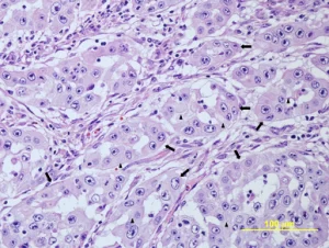
A lacrimal adenocarcinoma of the third eyelid in a 5-year-old Draft Horse was diagnosed based on histopathology. 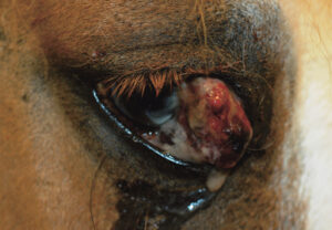
A 5-year-old Draft Horse gelding presented for evaluation of a large, fleshy, ulcerated third eyelid mass OD of 3 weeks duration.
Keywords: lacrimal adenocarcinoma horse, third eyelid tumor horse, equine ocular neoplasia, horse eye tumor case report, equine lacrimal gland adenocarcinoma, exenteration surgery horse eye, primary ocular tumor horse, equine ophthalmology oncology, third eyelid mass equine, demodex caballi horse eyelid
1Animal Eye Clinic, Westfield, Indiana, USA
2Department of Small Animal Medicine and Surgery, College of Veterinary Medicine, University of Georgia, Athens, GA, USA
Correspondence:
Rachel L. Davis, DVM, MS, Diplomate, ACVO – Ophthalmologist
Animal Eye Clinic
4750 Killarney Drive
Carmel, IN 46033
Email: info@indyaec.com