Proptosis – Canine
Rachel L. Davis1
VIN Publication
Abstract
Glaucoma refers to elevated intraocular pressure (IOP) that is either sustained or fluctuating. It can ultimately result in death of retinal ganglion cells. Glaucoma encompasses a broad category of diseases but is commonly referred to in the singular tense. Although normotensive glaucoma has been identified in people, it has not been documented in the cat or dog.
Learn about proptosis in dogs, an emergency condition where the eye is displaced from the orbit. Covers causes, diagnosis, treatment, surgery options, prognosis, and management in canine patients below.
Keywords: proptosis in dogs, canine globe proptosis, causes of proptosis in dogs, treatment for proptosis in dogs, enucleation for proptosis in dogs, surgical management of proptosis in dogs, globe displacement in dogs, orbital fractures and proptosis dogs, emergency treatment for eye trauma in dogs, prognosis of proptosis in dogs, proptosis in brachycephalic dogs, canine eye trauma emergency
Contributors:
1Revised by Rachel Davis DVM, MS, DACVO at Animal Eye Clinic, Westfield, Indiana, USA, on 5/04/2021
Original author was Ian P. Herring DVM, MS, DACVO, 4/16/2007
Correspondence:
Rachel L. Davis, DVM, MS, Diplomate, ACVO – Ophthalmologist
Animal Eye Clinic
4750 Killarney Drive
Carmel, IN 46033
Email: info@indyaec.com
Synonyms:
Globe proptosis
Disease Description:
Definition
Proptosis is sudden expulsion of the globe from the orbit causing the globe to be positioned anterior to the eyelids, with the eyelids entrapped behind the globe.
Etiology
Globe proptosis occurs with trauma (e.g. blunt, shearing, penetrating). A detailed history is important to determine the type of trauma sustained. The type helps to determine whether further evaluation is needed for systemic or other craniofacial trauma. In some cases, the nature of the trauma may be inferred but is not actually known. Proptosis may occur with minimal trauma in brachycephalic dogs because of their shallow orbits and large palpebral fissures. It is commonly associated with a dog fight or big dog-little dog altercation.1 Proptosis can also occur with choke injuries and overzealous restraint of brachycephalic dogs.
Orbital trauma is typically significant in mesocephalic and dolichocephalic dogs with proptosis because their orbits are larger and the globes are deep set.2 Similarly the feline orbit is more enclosed with bone than the canine orbit, so severe trauma is often required to cause proptosis.1,2
Diagnosis
Ophthalmic Examination Findings: Diagnosis is straightforward and made upon identification of the globe being located anterior to the orbit, with the eyelids entrapped behind it (Figure 1). Concurrent mild to severe orbital or head trauma may be present, with periocular subcutaneous emphysema, soft tissue swelling, skin lacerations or palpable skull fractures. When assessing a proptosed globe, it is important to assess both the periocular structures and globe viability (see Prognosis section below).
Physical Examination Findings: Because globe proptosis develops secondary to trauma, a thorough physical examination is performed to assess for systemic signs of trauma. In cats and non-brachycephalic dogs, evaluation for concurrent skull fractures, subcutaneous emphysema, epistaxis, periocular tissue damage, neurologic deficits, and systemic trauma is warranted.
Other Tests: Skull and thoracic radiographs and/or computed tomography may be indicated if facial fractures or thoracic trauma are suspected. Preoperative laboratory tests are recommended if systemic trauma has occurred.
Disease Description in This Species:
Signalment
Brachycephalic dogs are at higher risk of globe proptosis secondary to their conformational exophthalmos, relatively shallow orbit, globe exposure, and macropalpebral fissure.2 In one study of 29 dogs, proptosis occurred in 14 brachycephalic dogs.3 Another study including 43 dogs showed that brachycephalic dogs comprised 70% of the proptosis cases. Young[RD1] dogs (mean age 5.2 yrs) are at higher risk of globe proptosis.3 Dogs are typically presented on an emergency basis, often after a dog fight or sustaining head trauma. In a recent study, intact males were at higher risk for proptosis, due to fighting behavior. [RD2] The same study also reported that 56% of the proptosis cases were a result of dog fighting. Because the appearance of a proptosed globe is somewhat disturbing, most clients are distraught when they present their pet to the veterinary clinic.
Clinical Signs
The globe is positioned anterior to eyelids and orbit. The eye may be blind, with absent menace and pupillary light reflexes. Palpebral reflexes are often absent secondary to entrapment of eyelids posterior to globe. Chemosis, subconjunctival hemorrhage, bruising of the eyelids, hyphema, corneal ulceration secondary to exposure, and corneal drying (Figure 2) are all possible. The globe may be deviated (usually laterally) from avulsion of the medial extraocular muscles. The optic nerve may be severed and, in some cases, can be visualized externally (Figure 3). Periocular swelling, periorbital lacerations, subcutaneous emphysema, skull and mandibular fractures, epistaxis, and other systemic signs of trauma may be present. With severe head trauma alterations in mentation and other cranial nerves may be noted.
Etiology:
Bite wound
Iatrogenic
Trauma
Breed / Species Predilection:
Brachycephalic breeds
Sex Predilection:
None
Age Predilection:
Young adult
| Diagnostic Procedures: | Diagnostic Results: | |
| Radiography of head/skull | Facial fracture | |
| Computed tomography | Skull fracture/s | |
| Ocular examination | Cataract, lens opacity | |
| Corneal fluorescein staining positive | ||
| Excessive exposure of sclera | ||
| Eyelid, conjunctival laceration | ||
| Hyphema, blood anterior chamber eye |
Images:
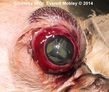
Figure 1. Proptosed eye (photograph)
Click here to see board discussion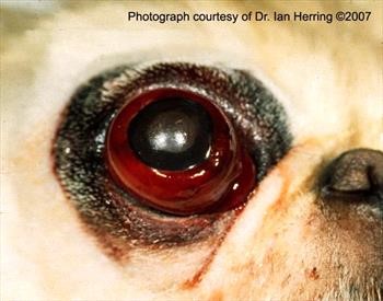
Figure 2. Globe proptosis in a 4-year-old Pekingese
Severe subconjunctival hemorrhage is evident, but there is no hyphema. Globe replacement was successful in achieving a cosmetic eye, although it remained blind. Courtesy of Dr. Ian Herring.
Figure 3. Proptosis with avulsion and exposure of the optic nerve. Click here to see board discussion 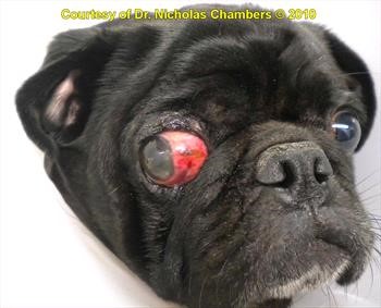
Figure 4. Post-proptosis OD with avulsion of the medial rectus muscle
Click here to see board discussion
Figure 5. Temporary tarsorrhaphy for proptosis.
Click here to see board discussion
Figure 6. Severe bilateral proptosis in an 11-year-old Boston terrier. Due to avulsion of most extraocular muscles in this case, enucleation was the only option. The source of trauma was unknown, but the severity at presentation was, in part, due to self trauma. Courtesy of Dr. Ian Herring.
Treatment / Management:
SPECIFIC THERAPY
Damage to the globe, optic nerve, and periocular structures is sustained at the time of the trauma, thus immediate replacement of the globe after presentation may not improve prognosis for vision. However, prompt replacement of the globe helps decrease the incidence of corneal ulceration and prevents severe swelling of the conjunctiva and eyelids from prolonged decreased venous return.4 Globe proptosis is considered a true ophthalmic emergency. The globe should be replaced as soon as possible if it is considered a good candidate for retention and the dog can undergo anesthesia.
Preoperative Medical Therapy
Upon initial presentation the cornea can be protected with a viscous lubricant (e.g. artificial tear ointment) to help avoid ulceration during ophthalmic and physical examination.
Preoperative Assessment
Although the patient is not able to close the palpebral fissure, a positive menace or dazzle response in a visual eye may be detected by movement of the eyelids or movement of the head away from the stimulus. Additionally, an indirect pupillary light response (PLR) from the proptosed eye to the unaffected eye may be used to assess for viability of vision. A recent study also concluded that the indirect PLR is a significant prognositic indicator of vision after globe reduction. Even[RD3] if the globe is not deemed viable, replacement may be attempted if the periorbital and periocular tissues appear intact.
Because the medial rectus muscle is the shortest extraocular muscle, this muscle is often damaged or transected during globe proptosis.1 Thus, permanent lateral strabismus may occur after avulsion of the medial rectus muscle (Figure 4). Permanent strabismus may cause increased corneal exposure or poor eyelid closure, which resultant chronic corneal ulceration or keratitis, or corneal infection with stromal melting. These sequelae often necessitate enucleation at a later date. Prior to surgery the client should be warned of the possibility of permanent strabismus; decreased ocular motility; absence of vision; and/or the need for enucleation of the globe later.
Surgical Reduction and Tarsorrhaphy
Globe reduction is performed with the patient under general anesthesia. Local anesthetic retrobulbar blocks are not recommended as they may cause further orbital and periocular tissue swelling. After induction of anesthesia, the periocular fur is gently clipped and cleaned with dilute betadine (1:10) solution. The corneal surface is generously lavaged and lubricated.
After cleaning, strabismus hooks may be inserted under the eyelids to manipulate them anteriorly and away from the globe so they may return to a normal position.1 Because most clinics do not have strabismus hooks readily available, towel clamps or Allis tissue forceps may be used to grasp the dorsal and ventral central eyelids as close to the margin as possible without damaging the globe or conjunctiva. While the eyelids are manipulated gentle manual pressure is concurrently applied to the cornea, usually by an assistant. Alternatively, the horizontal mattress sutures used to complete the tarsorrhaphy can be preplaced. As the sutures are gently pulled together and the eyelids are everted, gentle pressure can be applied to the globe. A lateral canthotomy may be performed if there is significant swelling or if the globe is difficult to reduce manually.1
After reduction, a temporary tarsorrhaphy is performed that closes the central and lateral palpebral fissure.5 Horizontal mattress sutures of 3-0 to 5-0 nonabsorbable sutures are placed through stents (e.g. rubber bands, IV tubing) to prevent the sutures from pulling through the eyelids (Figure 5). The suture is passed into the free edge of the eyelid margin through the meibomian gland openings in order to prevent the suture from rubbing on the cornea.6 A small opening is left medially to allow for application of topical medication.
Note that placement of a third eyelid flap is not recommended because it does not provide enough structural support to encourage the globe to return to a normal position. It is also difficult to perform in brachycephalic dogs because it is typically not long enough to cover the displaced globe.
Enucleation
Globes that are severely deviated from avulsion of multiple extraocular muscles or have avulsion of the optic nerve should be enucleated (Figure 6).2 Enucleation may also be considered for proptosed eyes that have experienced penetrating trauma or severe hyphema. If the patient is unable to undergo anesthesia immediately because of head, facial or systemic trauma, enucleation may be performed once the animal is stable. If the globe is enucleated, accessory adnexal structures (i.e. eyelid margins, third eyelid, conjunctiva) are carefully excised to avoid complications from residual adnexal tissue.7
SUPPORTIVE THERAPY
After reduction/replacement of a proptosed globe, systemic anti-inflammatory (e.g. carprofen 2.2 mg/kg PO q 12 hrs) and analgesic (e.g. gabapentin 10-20mg/kg PO BID-TID[RD4] ) therapy is recommended for 5-7 days. Oral antibiotic therapy may also be warranted depending on the amount of soft tissue injury sustained during the trauma.1 Topical antibiotics (e.g. triple antibiotic ointment or solution q 8 hrs) and artificial tear ointment q 8 hrs are recommended for 10-14 days postoperatively. Topical steroids are not recommended after globe reduction as they may potentiate corneal melting if an ulcer occurs secondary to proptosis or reduction surgery. Supportive therapy of other injuries is instituted as needed.
MONITORING
After globe reduction, the patient is re-evaluated in 5-7 days. If the globe has returned to a normal position and all eyelid/periocular swelling has resolved or if complications are occurring (e.g. purulent discharge, persistent pain), the central tarsorrhaphy suture may be removed. Ideally, the tarsorrhaphy should remain in place for 10-14 days to allow complete healing of the periocular and orbital tissues.
Globe motility and position are assessed after suture removal, along with the presence of vision, hyphema, and corneal ulceration. Quantitative tear assessment is performed because keratoconjunctivitis sicca may be a complication.2 Other known complications include blindness, strabismus (especially exotropia or lateral strabismus), exposure keratitis, lagophthalmos, and glaucoma. Long-term periodic evaluation is warranted.2 A second surgery may be considered for eyes with persistent deviation of the globe and exposure keratitis. The globe may be partially rotated medially with nonabsorbable suture and a permanent medial canthoplasty performed. Globes causing recurrent or ongoing discomfort to the patient should be enucleated.
PROGNOSIS
At initial presentation prognosis for vision is more favorable if a direct pupillary light response is present; the eye is visual; the fundus appears normal; and an indirect pupillary light response to the contralateral eye is present.1 Prognosis for salvage of the globe is favorable if the visual indicators previously mentioned are present; if no extraocular muscle damage is present; if <2 extraocular muscles are avulsed; and there is minimal or no intraocular hemorrhage detected.1,2 Pupillary size at presentation is of no prognostic value. Poor prognostic indicators for retention of the globe include proptosis in a mesocephalic and dolichocephalic breed, hyphema, optic nerve damage, concurrent facial fractures or avulsion of ≥2 extraocular muscles.2 The prognosis for vision ranges from 20-41% in various studies. Vision was present in 28% of dogs in a study that assessed only results from globes that were reduced, excluding eyes that were enucleated or cases in which the patient was euthanized. This study also showed that brachycephalic dogs have a higher likelihood of vision after reduction, which would be reasonable since less force/trauma is required to proptose a brachycephalic dog’s eye. Patients that were assessed by and operated on by a veterinary ophthalmologist had a significant better prognosis. Lastly, as the first study to assess client satisfaction, this study showed that even if globes were not visual or became phthiscal after reduction, the client satisfaction was very high 8.8/10 and 7.8/10 respectively. Thus, even if a globe is not expected to be visual, reduction should be considered if the globe is deemed salvageable. [RD5]
Special Considerations:
Other Resources:
Recent VIN Message Board discussions on proptosis
Client Handout on eye injuries
Proceedings articles that discuss proptosis
For more images of proptosis and its complications see the slideshow in the Image Library
Ophthalmology Fun Case 48
Ophthalmology Fun Case 57
Differential Diagnosis:
Exophthalmos
Orbital fractures with periocular swelling
- Spiess BM, Gelatt KN : Diseases and Surgery of the Canine Orbit. Veterinary Ophthalmology, 4th ed. Wiley-Blackwell, Oxford UK pp. 547-49.
- Gilger BC, Hamilton HL, Wilkie DA, et al: Traumatic ocular proptoses in dogs and cats: 84 cases (1980-1993). J Am Vet Med Assoc 1995 Vol 206 (8) pp. 1186-1190.
- Fritsche J, Ruhli M, Spiess B, et al: [Prolapse of the eyeball in small animals: a retrospective study of 36 cases]. Tierarztl Prax 1996 Vol 24 pp. 55-61.
- Mandell DC: Ophthalmic emergencies. Clin Tech Small Anim Pract 2000 Vol 15 (2) pp. 94-100.
- Cho J: Surgery of the globe and orbit. Top Companion Anim Med 2008 Vol 23 (1) pp. 23-37.
- Stades FC, Gelatt KN, Gelatt KN: Diseases and Surgery of the Canine Eyelid. Veterinary Ophthalmology, 4th ed. Wiley-Blackwell, Oxford UK pp. 612.
- Ward AA, Neaderl MH: Complications from residual adnexal structures following enucleation in three dogs. J Am Vet Med Assoc 2011 Vol 239 (12) pp. 1580-30.