Establishing a reproducible method for the culture of primary equine corneal cells
Rachel L. Davis1,2 | Ursula M. Dietrich2 | Thomas M. Krunkosky3 | David J. Hurley4 | Adrian J. Reber4
Abstract
Objective
To establish a reproducible method for the culture of primary equine corneal epithelial cells, keratocytes, and endothelial cells and to describe each cell’s morphologic characteristics, immunocytochemical staining properties and conditions required for cryopreservation.
Procedures
All three equine corneal cell types were successfully grown in culture. Cultured corneal endothelial cells were large, hexagonal cells with a moderate growth rate. Keratocytes were small, spindloid cells that grew rapidly. Epithelial cells had heterogenous morphology and grew slowly. The endothelial cells and keratocytes stained positive for vimentin and were morphologically distinguishable from one another. The epithelial cells stained positive for cytokeratin. Keratocytes and endothelial cells were able to be cryopreserved and recovered. The cryopreserved cells maintained their morphological and immunocytochemical features after cryopreservation and recovery.
Results
This work establishes reproducible methods for isolation and culture of equine corneal keratocytes and endothelial cells. Cell morphology and cytoskeletal element expression for equine corneal epithelial cells, keratocytes, and endothelial cells are also described. This has not previously been reported for equine corneal cells. This report also demonstrates the ability to preserve equine keratocytes and endothelial cells for extended periods of time and utilize them long after the primary-cell collection, a feature that has not been reported for veterinary corneal cell culture.
Discussion
Intravitreal dexamethasone injections in dogs with ocular blastomycosis is a simple procedure that significantly reduced the need for enucleation of eyes affected with ocular blastomycosis. This procedure may help prevent loss of eyes in dogs affected with ocular blastomycosis. None.
Images
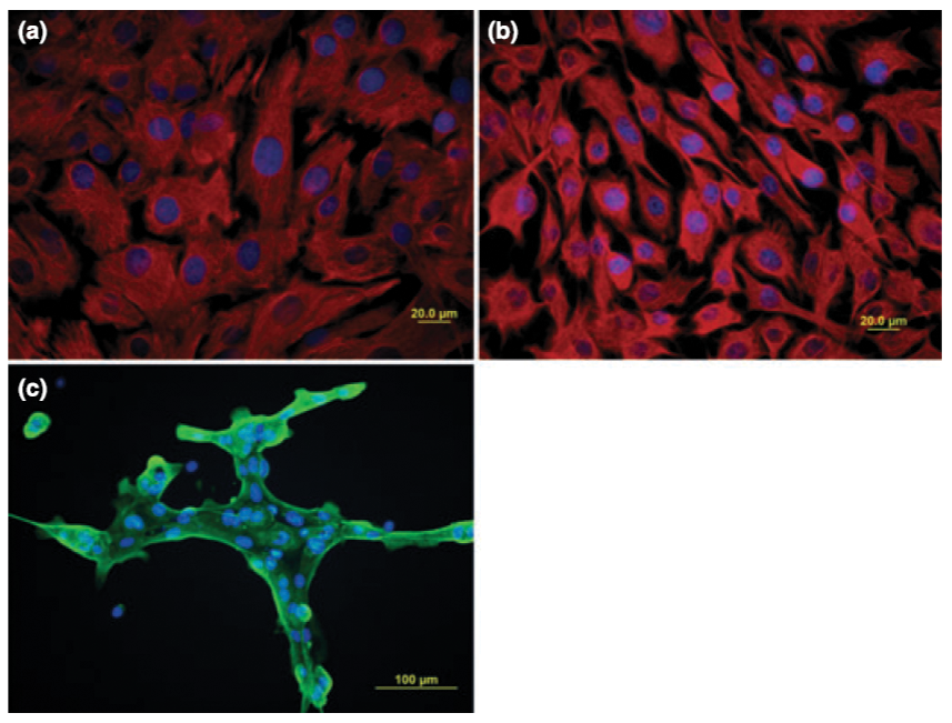
Figure 2. Photomicrographs of primary equine
corneal cells plated and fixed in glass slide chambers for immunohistochemical staining. Primary equine corneal endothelial cells (a) and keratocytes (b) depicted at ·400 magnification stained positive with anti-vimentin antibody CY3. Vimentin positive cells appear red with DAPI-stained nuclei appearing blue. Primary equine corneal epithelial cells stained positive
with high molecular-weight anti-cytokeratin
antibody 34BE12 and wide molecular-weight range
anti-cytokeratin antibody ST1 (c). These cells clustered in culture, as represented at ·200 magnification. A representative culture stained with antibody ST1 is depicted. Cytokeratin positive cells appear green with DAPI-stained nuclei appearing blue.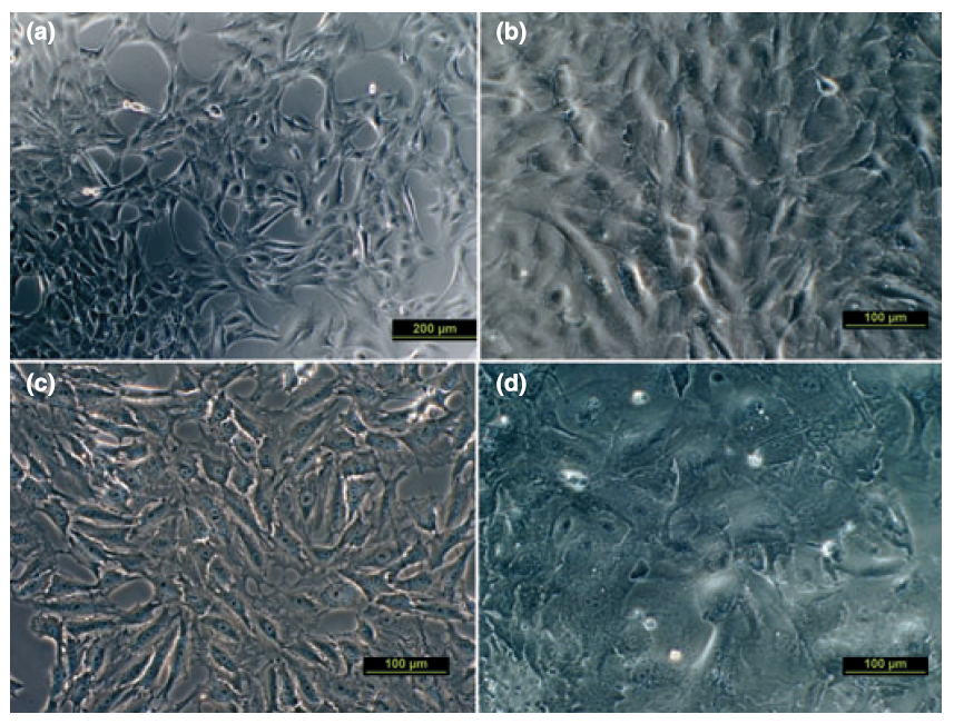
Figure 1. Photomicrographs of primary equine
corneal cells plated in glass chamber slides for
morphologic evaluation. When subconfluent,
endothelial cells demonstrated long cytoplasmic
processes and markedly flattened morphology. A
representative culture photographed at ·100
magnification is depicted (a). At confluency,
endothelial cells assumed a polygonal shape with
cobblestone-like appearance (b). Keratocytes were
small, spindloid cells with homogenous orphology
exhibiting a rapid growth rate (c). The epithelial
cells were large, polygonal cells with heterologous
morphology that readily formed clusters (d).
Confluent endothelial cells (b), keratocytes (c), and
epithelial cells (d) are photographed here at ·200
magnification.
Keywords: equine corneal cell culture, horse corneal epithelial cells, equine keratocyte culture, equine corneal endothelial cells, veterinary ophthalmology corneal research, cryopreservation equine corneal cells, immunocytochemical staining horse cornea, primary corneal cell isolation horses, equine corneal morphology study, veterinary corneal cell preservation
1Animal Eye Clinic, Westfield, Indiana, USA
2Department of Small Animal Medicine and Surgery, College of Veterinary Medicine, University of Georgia, Athens, GA USA
3Department of Anatomy and Radiology, College of Veterinary Medicine, University of Georgia, Athens, GA 30602, USA
5Department of Population Health, College of
Veterinary Medicine, University of Georgia, Athens, GA 30602, USA
Correspondence:
Rachel L. Davis, DVM, MS, Diplomate, ACVO – Ophthalmologist
Animal Eye Clinic
4750 Killarney Drive
Carmel, IN 46033
Email: info@indyaec.com