Chronic Superficial Keratitis (CSK) – Canine
Rachel L. (Mathes) Davis1
Veterinary Information Network (VIN)
Abstract
Chronic superficial keratitis (CSK), or pannus, is a bilateral disorder of the canine cornea characterized by progressive, often raised, corneal pigmentation, vascularization and hazy opacification (white or grayish).1 The lesions are not painful and are seen as small or large areas of red, brown or gray corneal discoloration typically arising from the ventrotemporal or temporal limbus (see images). The lesions may have a leading edge of white lipid deposits. In approximately 70% of cases, the third eyelid is concurrently affected (termed plasmacytoma) having a thickened, hyperemic margin with multifocal depigmentation and a raised irregular conjunctival surface.2
Chronic superficial keratitis (pannus) is an immune-mediated corneal disease in dogs that causes progressive pigmentation, vascularization, and haze. Learn about causes, breed predisposition, diagnosis, treatment with topical immunosuppressants, UV light effects, and long-term management below.
Keywords: chronic superficial keratitis, pannus, CSK, pannus in dogs, corneal pigmentation dogs, canine pannus, immune-mediated eye disease dogs, german shepherd eye disease, corneal vascularization dogs, canine corneal inflammation, UV light eye disease dogs, cyclosporine pannus therapy, tacrolimus pannus treatment, canine ophthalmology, chronic keratitis dogs, autoimmune eye disease dogs
Contributors:
1Rachel Davis DVM, MS, DACVO at Animal Eye Clinic, Westfield, Indiana, USA, June 2016
Correspondence:
Rachel L. Davis, DVM, MS, Diplomate, ACVO – Ophthalmologist
Animal Eye Clinic
4750 Killarney Drive
Carmel, IN 46033
Email: info@indyaec.com
Disease Description
Chronic superficial keratitis (CSK), or pannus, is a bilateral disorder of the canine cornea characterized by progressive, often raised, corneal pigmentation, vascularization and hazy opacification (white or grayish).1 The lesions are not painful and are seen as small or large areas of red, brown or gray corneal discoloration typically arising from the ventrotemporal or temporal limbus (see images). The lesions may have a leading edge of white lipid deposits. In approximately 70% of cases, the third eyelid is concurrently affected (termed plasmacytoma) having a thickened, hyperemic margin with multifocal depigmentation and a raised irregular conjunctival surface.2
Etiology
This disease is likely immune-mediated with recent evidence suggesting an altered haplotype3 of major histocompatibility (MHC) class II or abnormal MHC class II expression4 as contributory in disease development. CD4 expressing lymphocytes are the predominant infiltrative cell type in CSK.5 There is a strong German Shepherd breed predilection, suggesting possible heritability of the condition.2,6,7 CSK has been shown to be mediated by increased UV light exposure, with altitude significantly affecting the prevalence of this disease.8 UV light increases expression of corneal matrix metalloproteinase-2 (MMP-2), an inflammatory mediator also shown to be increased in corneal cells from CSK patients, suggesting a possible link to disease development.9
Diagnosis and Ophthalmic Examination Findings
Diagnosis can usually be made based on the appearance of the corneal lesions in breeds typically affected by the disease. Because corneal pigmentation, vascularization and opacification may occur with chronic corneal irritation, other underlying factors such as keratoconjunctivitis sicca, trichiasis, distichia or ectopic cilia causing the corneal lesions should be ruled out. Pannus is not painful, helping to differentiate it from other causes of corneal pigmentation and vascularization.
Etiology
- Genetic, hereditary
- Idiopathic, unknown
- Immune-mediated disease
- Ultraviolet light
Breed Predisposition
- Australian Shepherd
- Belgian Tervuren
- Border Collie
- German Shepherd
- Greyhound
- Siberian Husky
Sex Predisposition
Some studies report a higher prevalence in male dogs (Chavkin, Drahovska) while another study reported a higher incidence in female dogs (Jokinen). There is likely not a true sex predisposition for the disease.
Age Predisposition
Dogs between 4-7 years
Clinical Signs
- Progressive corneal pigmentation, vascularization, haze and edema
- Usually emerging from the ventrotemporal or temporal limbus, although it may occur anywhere along the limbus and cornea
Diagnostic Procedures
Ophthalmic examination – corneal pigmentation, vascularization and edema, often raised corneal granulation and irregular lesions present
Cytology – lymphoplasmacytic inflammation
Images
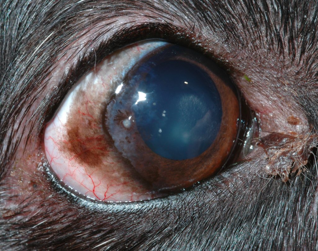
A clinical photograph depicting a patient affected with CSK shows infiltrative corneal pigmentation originating from the temporal limbus. There are admixed arborizing corneal vessels present. Photograph courtesy of Amy Baker, DVM, DACVO. 
A clinical photograph depicting a patient affected with CSK four weeks after instituting topical therapy shows multifocal pigment and fibrosis with admixed vascularization originating from the temporal and dorsal limbus. Note the multifocal thinning of pigment with the iris visible through the pigment in areas. Photograph courtesy of Amy Baker, DVM, DACVO.
Treatment/Management/Prognosis
Topical therapy is the mainstay of therapy for most cases of CSK. Topical cyclosporine,10,11 dexamethasone,10 tacrolimus in DMSO12 and pimecrolimus13 have been shown to significantly improve corneal CSK lesions and plasmacytic conjunctivitis. Typically, a combination of topical steroids (prednisolone acetate, dexamethasone or neopolydex) and immune-modulating therapy (tacrolimus or cyclosporine) are utilized initially at a 3-4x/day frequency then tapered to the lowest effective dose based on clinical response.1 Long term immune-modulating topical therapy is always needed to control the disease as it is progressive and will recur with discontinuation of therapy. Middle-aged to older dogs and dogs living in areas of the country with lower UV light levels typically respond well to topical therapy and may be managed long term with this alone.1
Radiotherapy using soft X-rays,14 strontium-90 therapy with or without superficial keratectomy15 and cryotherapy16 have been described to treat aggressive CSK cases refractory to medical therapy. These adjunctive therapies have been partially successful with concurrent with aggressive topical therapy, but recurrence tends to occur over time.
Prevention
There are no specific measures that may be taken to prevent CSK. Because CSK is mediated by UV light, decreased exposure to UV light is recommended for dogs affected with this disease. UV-blocking contact lenses were not shown to be helpful as an adjunctive therapy for dogs with CSK, although secondary corneal irritation from prolonged contact wear likely affected the results.17 Keeping affected patients inside during the day or UV protective eyewear (www.doggles.com) during outdoor activity may be recommended along with topical therapy. Studies are lacking on the effects of UV protective eye wear; however, anecdotally this seems to be beneficial for affected dogs spending extended periods of time outside (e.g. working and military dogs, dogs hiking in mountainous regions, etc.).
Differential Diagnosis
- Corneal granulation tissue
- Keratoconjunctivitis sicca
- Pigmentary keratitis/keratopathy
References
- Gilger, BC. Diseases and Surgery of the Canine Cornea and Sclera. Veterinary Ophthalmology 4th Pp 722-723.
- Drahovska, et al. A retrospective study of the occurrence of CSK in 308 GCDs: 1999-2010. Pol J Vet Sci 2014
- Jokinen, et al. MHC class II risk haplotype associated with canine CSK in GSD. Vet Immunol Immunopathol 2011
- Williams, DL. MHC class II expression in the normal canine cornea and in canine CSK. Vet Ophthalmol 2005
- Williams, DL. Histological and immunohistochemical evaluation of canine chronic superficial keratitis. Res Vet Sci. 1999
- Stanley, RG. Superficial stromal keratitis in the dog. Aust Vet J 1988
- Bedford, et al. Corneal pannus (chronic superficial keratitis) in the GSD. J Small Anim Pract 1979
- Chavkin, et al. Risk factors for development of CSK in dogs. JAVMA 1994
- Chandler, et al. Modulation of matrix metalloproteinases by UV radiation in the canine cornea. Vet Ophthalmol 2008
- Williams, et al. Comparison of topical cyclosporine and dexamethasone for the treatment of CSK in dogs. Vet Rec 1995
- Read, RA. Treatment of canine nictitans plasmacytic conjunctivitis with 0.2% CSA ointment. J Small Anim Pract 1995
- Balicki, I. Clinical study on the application of tacrolimus and DMSO in the treatment of chronic superficial keratitis in dogs. Pol J Vet Sci 2012
- Nell, et al. The effect of topical pimecrolimus on keratoconjunctivitis sicca and chronic superficial keratitis in dogs: results from an exploratory study. Vet Ophthalmol 2005
- Allgoewer, et al. Radiotherapy for canine chronic superficial keratitis using soft X-rays (15kV). Vet Ophthalmol 2010
- Hocht, et al. Treatment of keratitis superficialis chronica of the dog with strontium-90. Strahlenther Onkol (article in German) 2002
- Azoulay, T. Adjunctive cryotherapy for pigmentary keratitis in dogs: a study of 16 corneas. Vet Ophthalmol 2014
- Denk, et al. The effect of UV-blocking contact lenses as a therapy for canine chronic superficial keratitis. Vet Ophthalmol 2011