Published by Rachel Davis, DVM, MS, DACVO July 2017
Publication: Veterinary Information Network (VIN)
Disease Description
Persistent hyperplastic primary vitreous (PHPV), also known as persistent hyperplastic tunica vasculosa lentis (PHTVL) or PHPV/PHTVL, is a condition of the vitreous in which the ophthalmic embryologic vascular structures do not regress normally.1,2 Because the normal embryologic vitreal vasculature is intimately associated with the posterior lens surface/capsule and exits the eye at the posterior pole, abnormalities of these areas, especially the posterior lens, are also frequently seen in patients affected by PHPV.3.4 The degree of severity is variable ranging from an incidental finding of a retrolental fibrous strand5 to complete hyphema, severe ocular lens malformations, secondary glaucoma and patent, aberrant vasculature within the lens and vitreous.6 Clinically, patients affected with PHPV are typically presented for cataract evaluation, as most of these dogs have some level of lenticular involvement.
Etiology
PHPV is typically unilateral and non-inherited with sporadic appearance.7 In a small percentage of cases (~10%), the condition is bilateral and inherited, thought to be the result of inbreeding.7 These cases have been reported in the Doberman pinscher8,9 and Staffordshire bull terrier breeds.10,11 PHPV with retinal degeneration has been shown to be an autosomal recessive trait in Miniature Schnauzers,12 although retinal degeneration is typically not seen in other breeds with PHPV.
The underlying cause is not known, but has been suggested to be abnormal lens development,2,13 failure of of normal anti-angiogenic vitreal growth factor expression,14-16 failure of macrophagic-induced regression of the hyaloid vasculature17-19 or abnormal transformation of hyalocytes to phacocytically active cells for a short period of time during normal hyaloid vascular regression.20 Whatever the underlying etiology, the pathology becomes apparent after day 30 of canine embryologic development.16 While lenticular abnormalities are common in patients affected with PHPV,1,2,16,17 true lenticular developmental disease (lenticonus, microphakia, etc.) may occur independent of PHPV.16
Diagnosis
Ophthalmic Examination Findings
Because PHPV varies widely in severity of disease, the range of clinical ophthalmic findings is also wide. PHPV may be seen as an incidental white to gray retrolental strand in the vitreous on routine ophthalmic examination,5 although likely most of these cases go unidentified due to the location of the strand unless the patient is, for some reason (breeding exam, other ocular disease), being examined by a board-certified veterinary ophthalmologist. More commonly, these cases are affected with variable leukocoria and/or cataract formation, typically unilaterally.21-25 Patients with more severe PHPV’s and extensive vascularization may have complete hyphema, uveitis and/or secondary glaucoma.26 Depending on the severity of the ocular changes and whether the patient is affected uni- or bilaterally, decreased vision or blindness may be present.
Physical Examination Findings
PHPV is a primary ophthalmic condition and does not cause any systemic changes, although patients may have behavioral changes related to decreased vision or loss of vision.
Disease Description in this Species
Signalment
PHPV is a developmental disease, thus young dogs are typically affected.
Clinical Signs
Mild PHPV may be seen as an incidental white to gray retrolental strand in the vitreous on routine ophthalmic examination.5 More commonly, PHPV causes variable leukocoria and/or cataract formation, typically unilaterally, although it be bilateral.21-25 The leukocoria may appear as a wedge-shaped or triangular posterior lenticular cataract, a hemorrhagic area within the posterior lens or a white capsular opacity of the posterior lens.22,23 The PHPV may not be readily apparent based on the clinical examination alone because it is obscured by the lens opacification/s, requiring an ultrasonographic diagnosis.22,26,27 Patients with more severe PHPV’s and extensive vascularization may have complete hyphema, uveitis and/or secondary glaucoma.26 In Miniature Schauzers, PHPV may occur with retinal degeneration concurrently.12 A single case of a PHPV suspected of leading to a lens capsular rupture and secondary intraocular sarcoma has been reported in a Golden retriever.28 Depending on the severity of the ocular changes and whether the patient is affected uni- or bilaterally, decreased vision or blindness may be present.
Etiology
Non-inherited
Inherited
Genetic
Embryologic
Breed Predilection
Doberman pinscher
Staffordshire bull terrier
Miniature schnauzer
Bloodhound
Siberian husky
Basset hound
Mixed
Spanish pachon
Samoyed
Golden retriever
Bouvier des Flandres29
Sex Predilection
None
Age Predilection
Young dogs
Diagnostic Procedures
Ophthalmic examination
Ocular ultrasound (with and without Doppler)
Electroretinogram
Images

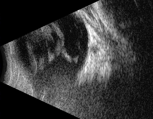
Figure 1: A curvilinear hyperechoic vitreal opacity may be seen centrally extending from the posterior lens to the posterior pole of the globe. There is concurrent hyperechogenicity of the lens, consistent with a complete cataract seen clinically, and lenticonus as seen by the posterior outpocketing of the lens and abnormal wedge-shape of the posterior lens. This patient underwent routine phacoemulsification of the cataract with intraocular lens implantation.

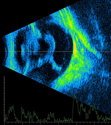
Figure 2: A Doppler ultrasonic image of the patient depicted in figure 1 shows no active blood flow through the PHPV. This can be helpful information prior to cataract surgery in assessing the overall risk of intravitreal hemorrhage intraoperatively. This patient underwent routine phacoemulsification of the cataract with intraocular lens implantation.
Treatment/Management/Prognosis
Specific Therapy
Treatment of PHPV depends on the severity of the condition and whether it affects patient vision or not. A PHPV found incidentally with with lenticular abnormalities or a PHPV causing small posterior cataracts with no visual disturbances does not require treatment. PHPV causing larger posterior lenticular opacities affecting vision may be referred to a board-certified veterinary ophthalmologist for cataract surgery to restore normal vision. In some cases, the PHPV causes complete cataract formation and the patient may be referred for cataract evaluation/surgery (in which case the PHPV is identified during the preoperative ocular ultrasound). Doppler is helpful preoperatively to assess overall risk of intravitreal hemorrhage intraoperatively. Most cases with cataract formation and PHPV can be successfully treated with phacoemulsification +/- planned posterior capsulorrhexis and intraocular lens implantation. In some cases with severe lenticonus or a large capsulorrhexis, an intraocular lens cannot be implanted; however, these patients typically maintain functional vision left aphakic. In cases with severe intraocular disease attributable to PHPV such as hyphema, uveitis or secondary glaucoma, enucleation may be the best option for long-term comfort.
Supportive Therapy
If a large cataract is present, topical steroidal (e.g. prednisolone acetate, dexamethasone) or non-steroidal (e.g. flurbiprofen, diclofenac) therapy may be instituted SID-BID for secondary lens-induced uveitis.
Monitoring and Prognosis
For most cases of PHPV affected with cataracts, the prognosis is excellent to good with cataract extraction (phacoemulsification) and intraocular lens implantation. In cases with cataract formation and intralenticular hemorrhage, the prognosis is good to guarded with surgery, although intraocular or intravitreal hemorrhage may be encountered intraoperatively. If the PHPV is an incidental retrolental strand, the prognosis is excellent. If the PHPV is very extensive, causing major intraocular pathology or secondary glaucoma and the eye is painful, the prognosis is poor to grave and enucleation should be pursued.
In Doberman pinschers, the incidence of severely affected dogs decreased from 19% to 8% from 1978-1987 utilizing selective breeding.30-32 Selective breeding against dogs affected with bilateral, inherited PHPV should be utilized for the Doberman pinscher, Staffordshire bull terrier and Miniature Schnauzer breeds.
Differential Diagnosis
Other causes of leukocoria
Lenticonus
Vitreal hemorrhage
Vitritis/fibrin
Vitreal degeneration/opacification
Cataract
References
- Davidson MG & Nelms SR. Diseases of the lens and cataract formation. In: Gelatt KN, ed. Veterinary Ophthalmology. 1999; 3rd ed. Philadelphia: Lippincott William & Wilkins. pp:797–825
- Boeve MH et al. Persistent hyperplastic tunica vasculosa lentis and primary vitreous (PHTVL/PHPV) in the dog: a comparative review. Prog Vet Comp Ophthalmol. 1992;2:163–172
- Boevé MH, van der Linde-Sipman JS, Stades FC, Vrensen GF. Early morphogenesis of persistent hyperplastic tunica vasculosa lentis and primary vitreous. A transmission electron microscopic study. Invest Ophthalmol Vis Sci.1990 Sep;31(9):1886-94.
- Boevé MH, van der Linde-Sipman JS, Stades FC. Early morphogenesis of the canine lens capsule, tunica vasculosa lentis posterior, and anterior vitreous body. A transmission electron microscopic study. Graefes Arch Clin Exp Ophthalmol.1989;227(6):589-94.
- Boroffka SA, Verbruggen AM, Boevé MH, Stades FC. Ultrasonographic diagnosis of persistent hyperplastic tunica vasculosa lentis/persistent hyperplastic primary vitreous in two dogs. Vet Radiol Ultrasound.1998 Sep-Oct;39(5):440-4.
- Bayón A, Tovar MC, Fernández del Palacio MJ, Agut A. Ocular complications of persistent hyperplastic primary vitreous in three dogs. Vet Ophthalmol.2001 Mar;4(1):35-40.
- Colitz CM, Malarkey DE, Woychik RP, Wilkinson JE. Persistent hyperplastic tunica vasculosa lentis and persistent hyperplastic primary vitreous in transgenic line TgN3261Rpw. Vet Pathol.2000 Sep;37(5):422-7.
- Stades FC (1983) Persistent hyperplastic tunica vasculosa lentis and persistent hyperplastic primary vitreous in Doberman pinschers: genetic aspects. J Am Anim Hosp Assoc19:957–964
- Stades FC (1980) Persistent hyperplastic tunica vasculosa lentis and persistent hyperplastic primary vitreous (PHTVL/PHPV) in 90 closely related Doberman pinschers: clinical aspects. J Am Anim Hosp Assoc 16:739–751
- Leon A et al(1986) Hereditary persistent hyperplastic primary vitreous in the Staffordshire bull terrier. J Am Anim Hosp Assoc22:765–774
- Curtis R, Barnett KC, Leon A. Persistent hyperplastic primary vitreous in the Staffordshire bull terrier. Vet Rec.1984 Oct 13;115(15):385.
- Grahn BH, Storey ES, McMillan C. Inherited retinal dysplasia and persistent hyperplastic primary vitreous in Miniature Schnauzer dogs. Vet Ophthalmol.2004 May-Jun;7(3):151-8.
- Spitznas M, Koch F, Pohl S: Ultrastructural pathology of anterior persistent hyperplastic primary vitreous. Graefe’s Arch Clin Exp Ophthalmol 228:487–496, 1990
- Lutty GA, Mello RJ, Chandler C, Fait C, Patz A: Regulation of cell growth by vitreous humour. J Cell Sci 76: 53–65, 1985
- Lutty GA, Thompson DC, Gallup JY, Mello RJ, Patz A, Fenselau A: Vitreous: An inhibitor of retinal extract-induced neovasculatization. Invest Ophthalmol Vis Sci 23: 52–56, 1983
- Boeve MH, van der Linde-Sipman T, Stades FC: Early morphogenesis of persistent hyperplastic tunica vasculosa lentis and primary vitreous, the dog as an otogenetic model. Invest Ophthalmol Vis Sci 29:1076–1086, 1988
- Cook CS: Embryogenesis of congenital eye malformations. Vet Comp Ophthalmol 5(2):109–123, 1995
- Jack RL: Regression of the hyaloid vascular system. An ultrastructural analysis. Am J Ophthalmol 74(2):261– 272, 1972
- Latker CH, Kuwabara T: Regression of the tunica vasculosa lentis in the postnatal rat. Invest Ophthalmol Vis Sci 21:689–699, 1981
- Balazs EA, Toth LZ, Ozanics V: Cytological studies on the developing vitreous as related to the hyaloid vessel system. Arch Ophthalmol 213:71–85, 1980
- Cullen CL, Grahn BH. Diagnostic ophthalmology. Persistent hyperplastic tunica vasculosa lentis and primary vitreous. Can Vet J.2004 May;45(5):433-4
- Verbruggen AM, Boroffka SA, Boevé MH, Stades FC. Persistent hyperplastic tunica vasculosa lentis and persistent hyaloid artery in a 2-year-old basset hound. Vet Q.1999 Apr;21(2):63-5.
- Boroffka SA, Verbruggen AM, Boevé MH, Stades FC. Ultrasonographic diagnosis of persistent hyperplastic tunica vasculosa lentis/persistent hyperplastic primary vitreous in two dogs. Vet Radiol Ultrasound.1998 Sep-Oct;39(5):440-4.
- Ori J, Yoshikai T, Yoshimura S, Takenaka S. Persistent hyperplastic primary vitreous (PHPV) in two Siberian husky dogs. J Vet Med Sci.1998 Feb;60(2):263-5.
- Gemensky-Metzler AJ, Wilkie DA. Surgical management and histologic and immunohistochemical features of a cataract and retrolental plaque secondary to persistent hyperplastic tunica vasculosa lentis/persistent hyperplastic primary vitreous (PHTVL/PHPV) in a Bloodhound puppy. Vet Ophthalmol.2004 Sep-Oct;7(5):369-75.
- Bayón A, Tovar MC, Fernández del Palacio MJ, Agut A. Ocular complications of persistent hyperplastic primary vitreous in three dogs. Vet Ophthalmol.2001 Mar;4(1):35-40.
- Boevé MH, Vrensen GF, Willekens BL, Stades FC, van der Linde-Sipman JS. Early morphogenesis of persistent hyperplastic tunica vasculosa lentis and primary vitreous (PHTVL/PHPV). Scanning electron microscopic observations. Graefes Arch Clin Exp Ophthalmol.1993;231(1):29-33.
- Graham KL, Krockenberger MB, Billson FM. Intraocular sarcoma associated with lens capsule rupture and persistent hyperplastic primary vitreous in a dog. Vet Ophthalmol.2016 Dec 23. doi: 10.1111/vop.12454. [Epub ahead of print]
- van Rensburg IB et al(1992) Multiple inherited eye anomalies including persistent hyperplastic tunica vasculosa lentis in Bouvier des Flandres. Prog Vet Comp Ophthalmol2:133–139.
- Stades FC, Boevé MH, van den Brom WE, van der Linde-Sipman JS. The incidence of PHTVL/PHPV in Doberman and the results of breeding rules. Vet Q.1991 Jan;13(1):24-9.
- Pfahler S,Menzel J,Brahm R, Rosenhagen CU, Hafemeister B, Schmidt U, Sinzinger W, Distl O. Prevalence and formation of primary cataracts and persistent hyperplastic tunica vasculosa lentis in the German Pinscher population in Germany. Vet Ophthalmol. 2015 Mar;18(2):135-40. doi: 10.1111/vop.12167. Epub 2014 Mar 27.
- Leppänen M, Mårtenson J, Mäki K. Results of ophthalmologic screening examinations of German Pinschers in Finland–a retrospective study. Vet Ophthalmol.2001 Sep;4(3):165-9.
